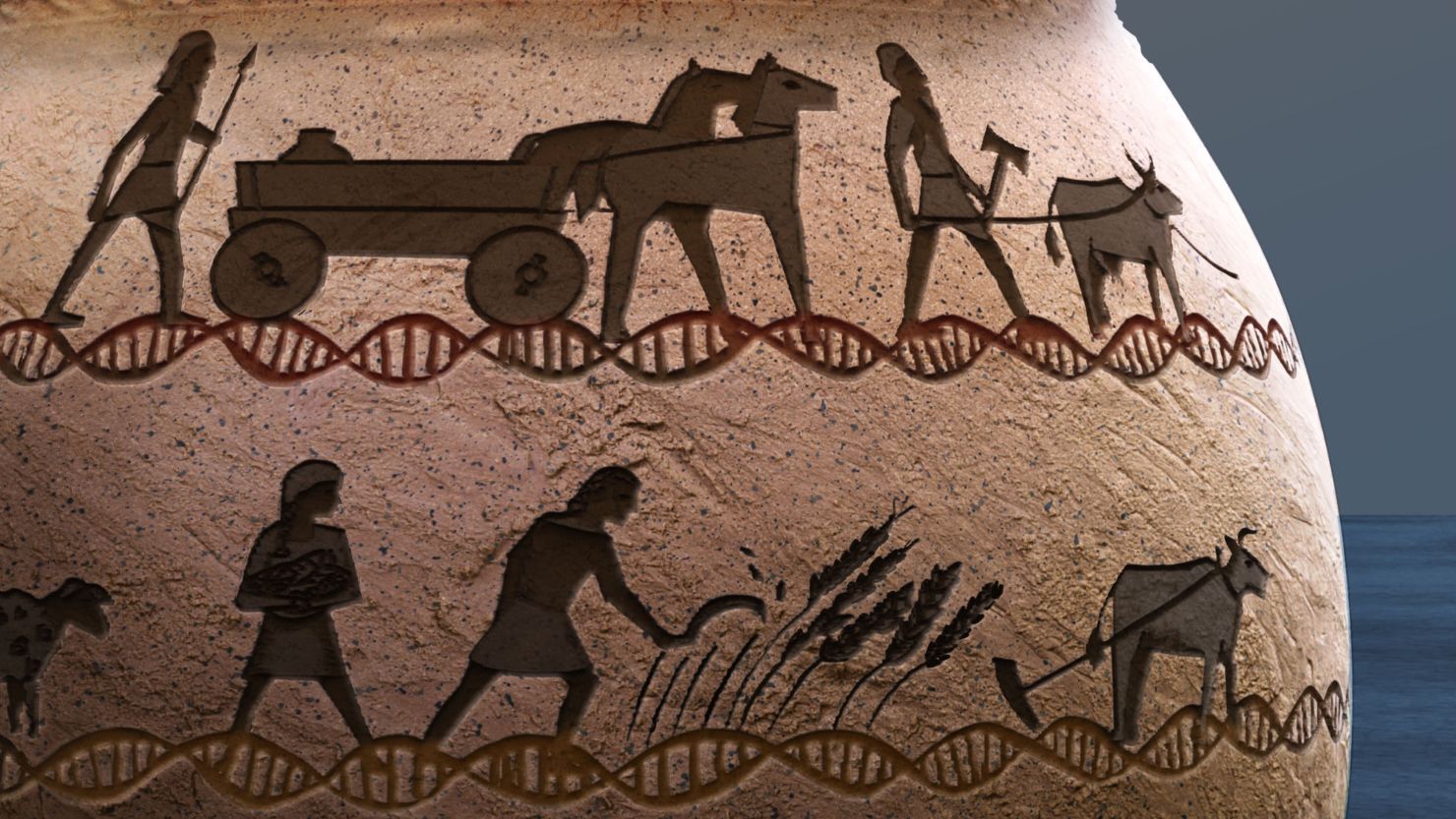
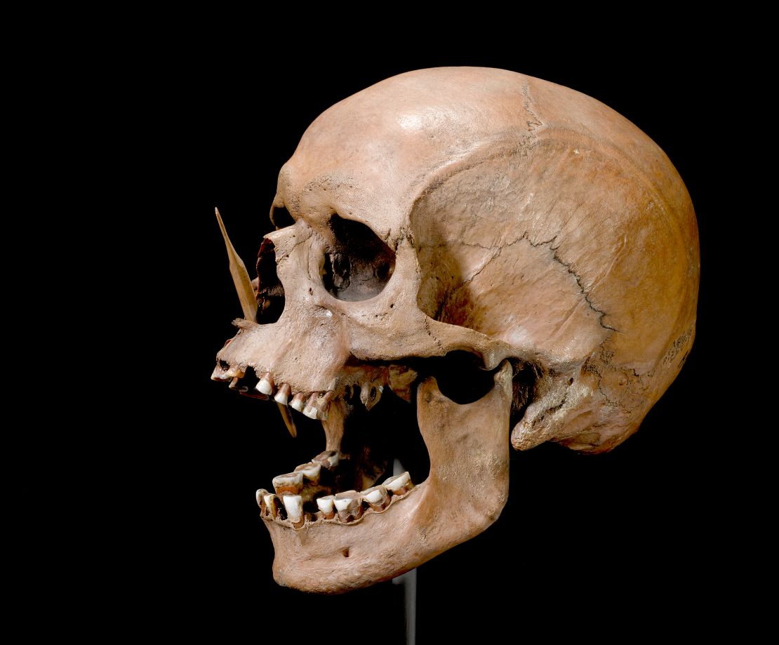
Fenbendazole

Fenbendazole Joe Tippens Protocol: A Simple Step-by-Step Guide

By Dr Frank Yap, MD – February 24, 2023

If you have been diagnosed with cancer, or have a loved one that is suffering from cancer, chances are you have heard about the Joe Tippens Cancer Protocol. The Fenbendazole Cancer Protocol has been gaining rapid interest over the past years following some fenbendazole advanced cancer success stories.

Joe Tippens founded the protocol after he was told a story about a scientist at Merck Animal Health that had been performing cancer research on mice. The research included injecting different types of cancers into different mice body parts. The scientists discovered, through trial and error, a product in their canine product line, fenbendazole, that was batting 1.000 in killing these different cancers in the mice. The scientist was later diagnosed with stage 4 brain cancer and was given a grim prognosis of only three months to live. She decided to try the fenbendazole, and after six weeks, showed a clean scan.

Joe Tippens had been initially diagnosed with small cell lung cancer. The cancer later spread to his neck, right lung, stomach, liver, bladder, pancreas and tail bone. Like the scientist from Merk Animal Hospital, Joe was told he only had three months to live. In 2017, after hearing the story of the scientist who treated her cancer with a canine drug, Joe decided he was going to do the same. However, in addition to taking the fenbendazole, Joe added his own ingredients to the regimen (curcumin, CBD oil, and vitamin E), thus creating the Joe Tippens Cancer Protocol.

Disclaimer: We in no way endorse the Joe Tippens Cancer Protocol as a cure for cancer, and as always, you should consult your physician before starting any new medications or treatment plans.

What Is Fenbendazole?

According to Wikipedia, Fenbendazole is a broad spectrum anthelmintic used against gastrointestinal parasites including: giardia, roundworms, hookworms, whipworms, the tapeworm genus Taenia (but not effective against Dipylidium caninum, a common dog tapeworm), pinworms, aelurostrongylus, paragonimiasis, strongyles, and strongyloides that can be administered to sheep, cattle, horses, fish, dogs, cats, rabbits, most reptiles, freshwater shrimp tanks as planaria and hydra treatments, as well as seals.

pastedGraphic.png

Common brand names include Pancur, Panacur C and Safe-Guard.

However, it is also being used by humans in a cancer treatment method known as the Joe Tippens Protocol.

Is fenbendazole a cancer cure scam? One study suggests that fenbendazole has “been safely utilized as an anti-parasitic for various different animal species and could be repurposed for treating human malignancies.” Other benzimidazoles, including albendazole, parbendazole, mebendazole and flubendazole have already been shown to have promising results in humans

Another study states that the “repurposing of veterinary drugs showing promising results for human use can result in considerable time and cost reduction required to develop new drugs.”

Fenbendazole Protocol – A Simple Step-by-Step Guide

The basic fenbendazole protocol people follow is surprisingly simple and includes a few added supplements to the fenbendazole:

** Mimi does not recommend CBD oil. It will scar the subtle energy bodies.

  1. Fenbendazole
  2. Curcumin
  3. CBD Oil
  4. Berberine
  5. Quercetin

1. Fenbendazole: One Packet a Day

Fenbendazole which has 222mg of Fenbendazole per gram: one packet of powder per day for seven days a week. It can be mixed with food such as yogurt or simply taken by itself. 

pastedGraphic.png

2. Curcumin: 600 mg a Day

600 mg per day of bioavailable curcumin, which is the active agent in the herb turmeric. Curcumin may help increase healthy p53 levels, and it has been shown to be a potentially effective cancer therapy supplement.

Click here to buy Theracurmin HP by Integrative Therapeutics (Amazon)

3. CBD Oil: 25 mg a Day

** Mimi does not recommend CBD oil. It will scar the subtle energy bodies.

25 milligrams daily, taken sublingually (under the tongue). The CBD oil should be high-purity level broad-spectrum. CBD has been shown to potentially modulate tumor growth.

Click here to buy medical-grade CBD Oil >> (use code RETHN2 for a 20% discount!)

A nurse’s tip: 

pastedGraphic_1.png
  • It is important to choose the right CBD for medical use: flower-derived, lab-tested, organic, whole-spectrum.
  • To enhance CBD healing response for cancer symptoms, slowly increase to 0.5ml of CBD twice daily for a total of 50 mg.

4. Berberine: 2-3 times a day

Berberine has shown important anti-tumor effects in numerous studies. These studies reported that Berberine could prevent the multiplication of cancer cells and inhibit metastasis and the spread of cancer cells. Berberine can work with Fenbendazole to further limit the cancer cells’ ability to take up glucose. This way, cancer cells are weakened and starved. 

Click here to buy Berberine (Amazon)

5. Quercetin: 1-2 a day

Due to its antioxidant, anti-tumor, and anti-inflammatory activities, quercetin has been studied extensively. Quercetin can inhibit the spread of many cancers such as prostate, cervical, lung, breast, and colon. Quercetin is not harmful to healthy cells yet powerful against cancer cells, making it a good candidate for a supplementary factor along with other anticancer medications.

Read More: Best Quercetin Supplements

FAQs

Can fenbendazole be taken in tablet, liquid or powder?

Fenbendazole is available in tablets (Happy Healing Store), liquid (Safeguard) and powder (Panacur). There may be other formulations available, but those are the primary products that patients are using.

pastedGraphic_2.png

Is it safe for humans to take fenbendazole?

Since fenbendazole is a veterinary medicine, and is not licensed for human use and there is no specific human safety data available. The similar human forms of this class of medications, mebendazole and albendazole, have been used for parasitic infections for many years and have a good safety record. Fenbendazole has been reported to have a very low degree of toxicity and a high degree of safety in animal experiments.

Is fenbendazole toxic for the liver?

Although fenbendazole is generally well tolerated, there have been reports of elevated liver enzymes from the use of fenbendazole, as well as the human medications mebendazole and albendazole. 

I’ve found a Fenbendazole brand which is not made by Merck, is it ok?

There are a variety of opinions on which product is best, but no research exists that compares the effectiveness of various formulations in humans. Merck and Safeguard are well established companies whose products have been safety tested in animals and used by veterinarians for many years. 

Would the fenbendazole protocol work with conventional treatment? – chemotherapy?

Yes, fenbendazole has the potential to synergize with conventional treatments, and possibly enhance treatment response. Some aspects of the protocol, such as vitamin E and curcumin, may interfere with chemotherapy and radiation due to their anti-oxidant properties. 

Some people take it 3 day on 4 off, some 1 gram a day, some 2 or even 4 grams a day. What is the correct dosage?

Joe Tippens, who popularized the use of Fenbendazole for cancer, recommends 222 mg for three consecutive days, then four days off. There are many people who take larger doses, more frequently. Since Fenbendazole is not licensed for the treatment of cancer in humans, there is no correct dose that is supported by research, pharmacists or physicians. The dose you choose to take should be assessed based on your other medications and treatments, your type of cancer, prognosis, organ function and lab values.

pastedGraphic_2.png

How long do I need to be on this protocol?

This will be up to you based on your treatment response to the protocol. It is reported that Joe Tippens continues to use the protocol for prevention.

How do I know when it is time to dose up?

This is an individual decision based on treatment response, tolerance, other treatments/medications and lab values. 

Q&A Session with Joe Tippens (Jan 2022)

Adam Payne

482 subscribers

Q&A Session with Joe Tippens

Share

More videos

Generative AI Is About To Reset Everything, And, Yes It Will Change Your Life | Forbes

Forbes • 344K views

21:25

CardioSmart | Living with Atrial Fibrillation (AFib)

CardioSmart • 346K views

55:03

Brad DeLong on Intellectual and Technical Progress (full) | Conversations with Tyler

Mercatus Center • 814 views

59:11

Penguins in Warm Weather | February 24, 2023

CNN 10 • 56K views

10:01

LIVE from Asbury University’s Spiritual Revival

CBN News • 141K views

2:35:10

ChatGPT Tutorial – A Crash Course on Chat GPT for Beginners

Adrian Twarog • 3.8M views

34:05

Alisa Childers Reflects on Visit to Asbury Revival

CBN News • 228K views

15:03

LISTEN LIVE: Supreme Court considers legal liability for big tech companies

PBS NewsHour • 96K views

2:52:10

🔊 How to use Audacity to Record & Edit Audio | Beginners Tutorial

Kevin Stratvert • 2.3M views

38:27

Flutter Forward 2023 Livestream

Flutter • 2M views

2:26:59

An Uncensored History of DARPA | Annie Jacobsen | Talks at Google

Talks at Google • 483K views

46:20

Washington Today (2-23-23): Transp. Sec. Buttigieg surveys toxic train derailment cleanup in Ohio

C-SPAN • 1.1K views

1:13:16

0:01 / 1:19:47

<div class=”player-unavailable”><h1 class=”message”>An error occurred.</h1><div class=”submessage”><a href=”https://www.youtube.com/watch?v=mnAX1-K81oQ” target=”_blank”>Try watching this video on www.youtube.com</a>, or enable JavaScript if it is disabled in your browser.</div></div>

What’s Joe Tippens doing in 2022 to stay cancer free?

One of the contributors from CancerDefeated.com met Joe in person and interviewed him in April 2022. Here’s what she discovered…

Joe’s scan was completely clear in early 2017. His scan in Feb. 2022 still shows no evidence of disease.

Since cancer is notorious for recurrences, Joe continues to follow his regimen for prevention… and plans to for the rest of his life. However, he recently revamped the rest of his entire cancer treatment protocol.

Joe has scaled back to three days a week on the FenBen, now that he’s been cancer-free for five years. He recommends staying on the seven-day fenbendazole protocol till you’re cancer-free for at least three years.

Based on his extensive research, he’s added things to his protocol. including Frankincense and berberine. All products are in a much more bioavailable form, too. He notes that getting absorption of nutrients into the bloodstream is a bigger problem than most people realize. His new protocol helps address this problem.

Joe spent much of 2021 collaborating with scientists and M.D.’s in the pharmaceutical and nutraceutical space, to develop processes that dramatically improve nutrient delivery into the bloodstream at previously unheard-of levels.

Here’s the three-pronged approach he uses in 2022 (R):

1: Support healthy cell division. Joe consumes cryogenically extracted CEM™ Hemp. Getting the terpenes and cannabinoids from full-spectrum hemp is critical to gaining its best benefits. The product is now cryo-extracted to preserve these substances. It also includes Frankincense and a Nano MCT oil.

2: Support healthy cell life cycles and clearance of dead cells with curcumin. Curcumin was always part of Joe’s protocol. He previously used the most bioavailable curcumin product he could find. He cringes when he relates how much he was depending on a nutrient that’s so 

Posted in Most Recent | 306 Comments

Reversing Chronic Kidney Disease with Niacin and Sodium Bicarbonate

TOWNSEND LETTER

The Examiner of Alternative MedicineSaturday, December 3, 2022

Reversing Chronic Kidney Disease with Niacin and Sodium Bicarbonate

By Stephen McConnell and W. Todd Penberthy
Orthomolecular Medicine News Service

Subscribe button

This story began with initial discovery, motivated by necessity. It would lead a few years later to reproducible documented reversal of chronic kidney disease (CKD) stages 1 or 2. Success was achieved using 3 to 5 cents/day of 100-500 mg niacin TID [three times a day] along with 1.0-1.8 grams of sodium bicarbonate (baking soda, 600 mg at lunch and 1.2 g before bed) with or without <2g/day elemental calcium, as calcium carbonate.

Excellent results from the use of niacin to treat CKD have now been documented for more than 25 case-studies. This approach is well supported by continuous basic and clinical research, including dozens of clinical trials that provide substantial evidence for the use of niacin and sodium bicarbonate. These approaches directly address the needs of the typical CKD patient. Unfortunately, this approach is rarely implemented in the clinical setting.

CKD commonly progresses with age as it is observed in 68% of Americans ≥60 years of age.1 Patients with CKD usually experience progressive loss of kidney function moving towards an increasing risk of end-stage renal disease (ESRD). CKD is the 9th leading cause of death in the US.2 Fortunately, there are several simple approaches including the addition of modest doses of niacin (immediate release- or IR-Niacin) that can reverse CKD in many patients as described here.

Approximately 786,000 people per year, in the US progress to ESRD (stage 5 CKD), which is generally considered an irreversible condition. Most of these become completely dependent on regular trips to dialysis. Estimation of the stages of CKD is based on (GFR) glomerular filtration rates starting with ≤60mL/1.7m2 for three months as definitive of initial CKD diagnosis. Unfortunately, a creatinine derived GFR (crGFR) is only as reliable as the serum creatinine measure. Use of this creatinine-based test has a “blind-area” in the earlier stages and frequently leads to an under-estimation of the true risk.


Stages of CKD

  1. Mild kidney damage, eGFR 90 or higher
  2. Mild loss of kidney function, eGFR 60-89
  3. Moderate loss of kidney function
    a. eGFR 45-59
    b. eGFR 30-44
  4. Severe loss of kidney function, eGFR 15-29
  5. Kidney failure or close to failure, eGFR less than 15


Niacin for CKD

Supplementation with daily low-dose niacin reliably reverses a large amount of the functional loss. This simple treatment is effective and critically important. Mortality rates with CKD are striking, as the five-year survival rate for patients doing long-term dialysis is 35% compared to 25% in those with diabetes [T2DM] in the USA.3

Routinely, the first treatment approaches utilized for CKD patients, in the later stages, generally targets control of dysglycemia and reduction of hyperphosphatemia according to KDIGO guidelines.4 Fortunately, there is an ever-increasing abundance of data revealing that simple niacin treatment is a profoundly effective treatment for reducing hyperphosphatemia—and that is just the beginning. In basic research the evidence in favor of niacin for CKD has continuously accumulated. Clinical research proves that the niacin stimulated pathways involving increased NAD synthesis, PCSK9 inhibition, sodium transporter effects, PPAR gamma activation, and more, are exceptionally well-suited to addressing CKD, multimorbidity, and ultimately all-cause mortality.5-41

The clinical and financial impact of CKD when it progresses to end-stage renal disease (dialysis-dependence; ESRD) is profound. Clinically, CKD progression quickly leads to lifelong dialysis with co-morbid life-threatening cardiovascular disease. Financially, the out-of-pocket cost of CKD is greater than cancer and stroke with ESRD dialysis costing 30.9 billion per year in 2013 or approximately 7.1% of total Medicare costs.42Medicare spends approximately $250,000/y for every CKD patient, prior to the transition to ESRD and dialysis. Annual costs per dialysis patient can range from $720k to $2.2m per year.43 These problems and their associated costs can be reduced by using 5 cents per day of niacin.

Originally, I (SM) was formally trained to operate a heart-lung machine, maintain full life-support and anesthesia, in the operating room monitoring patients undergoing open-heart surgery. Much later, I transitioned to working as a field scientist, MSL (Medical Science Liaison) in the advanced laboratory diagnostics industry. My primary clinical focus since that time has mainly been lipidology. Because of my initial education/training, addressing cardiovascular disease, I now focus on prevention: lipidology. This training gave me an appreciation for nicotinic acid (niacin, vitamin B3).

I have now personally observed more than 25 documented cases of individuals having their CKD progression not only halted but reversed with the addition of 3 to 5 cents worth of niacin, per day (with 1.8-2.4 g/day sodium-bicarbonate with/without 250-500 mg/day calcium-carbonate).


A Family Story

While I (SM) was learning lipidology in the period between 2002-2007, my father suddenly went to the ER late one Sunday night and my mother called me hysterically, “I took your father to the ER and now they are scheduling him for placement of stents.” I was concerned, as any son would be, but also as a scientist because I felt I may have ‘failed him,’ somehow: If only what I had already learned, I had only learned it, sooner.

My father was 81 at the time and he had been jumping rope for 30-minutes, twice daily. His body had a deceptively healthy look, and his triglycerides were low, but when we put it all together, he was “Pattern-B”—insulin resistant. He had always been a ‘stodgy,’ stubborn, stoic World War II veteran. He was very introverted and typically had a limited range of emotions: rage, laughter, and silence. Later, I would find out he had Asperger’s.

When I received the advanced laboratory data, it showed that he had low HDL2 and high ApoB. This is far more specific and confers much greater risk vs. an elevated LDL-C. Most importantly, this revealed he was insulin resistant (a.k.a. pre-diabetic). At the time, I really didn’t fully understand this. Even today, most clinicians really do not, due to continued reliance on using only tests for FBG and HbA1c. Ultimately, my father survived, and we continued to institute aggressive medical management: A hard lesson learned.

My father and my mother traveled everywhere together. They commuted, seasonally to Florida each winter, to escape the cold weather in Northwest Pennsylvania. On New Year’s Day, about six months after his MI and stent-placement procedure, I received a call from my mother, “Your father is in the hospital! They’re going to have to do open heart surgery!”

They needed to do an aortic-annuloplasty (aortic heart valve repair), in addition to a quintuple CABG (5 bypass grafts). I thought to myself, “this is getting worse and worse.” Having had previous personal experience working with thoracic surgeons during open-heart operations, I didn’t want the procedure to begin until my brother and I were able to be present. Fortunately, the young thoracic surgeon and the techniques planned were excellent.

Later, in the spring, they returned home to Erie, Pennsylvania, for the follow-up visit. Dr. Dave (the physician who asked me to set-up my 1st lipid clinic) said, “Hey I got some bad news for you. Your dad has renal insufficiency.” I said, “Oh my God, he’s in renal failure, what stage is he?” He did not know. That was a flag. Most clinicians don’t know what stage their CKD patients are because the lab doesn’t do calculations and the creatinine measure is not reliable or accurate. The creatinine measure has very little accuracy until after the CKD has ‘hit’ stage 3B, and beyond.44,45 So, a lot of these patients along the CKD disease continuum, through each progressive stage, appearing to have less risk vs. the ‘true’ risk that is present. It’s better to test a urine sample and see how much protein is recovered and run a Cystatin-C and a crGFR to calculate a more accurate value. At that time, I only knew he was in failure; but when I did the crGFR calculation, I could see that he was well into the latter portion, of CKD-stage 4.

Recently, I had been putting together a new treatment algorithm with substantial literature support, data, on CKD. I was lucky to have been mentored by Dr. William F. Finn.46 Even if a patient has not already been scheduled for dialysis, he explained, and especially if they are currently on dialysis, you must get the serum phosphorus down. Excessive phosphorous is toxic to the kidneys as well as virtually every organ system and the entire body.47,48 Phosphorus is a primary initiator of vascular calcification, among several other pathologies. If the kidneys start to lose a certain fraction of their normal function, the body can no longer efficiently clear phosphorous. When phosphorous serum levels reach abnormal levels, then you begin to saturate the tissues. Then phosphorous binds to calcium and it’s the phosphorous, not the calcium that starts the pathology leading to calcium phosphate stones.


Niacin Helps to Get the Phosphorous Down

Even after you bring serum phosphate down you still have it in the tissues. The only biomarker available in a clinical setting, Fibroblast Growth Factor-23 (FGF-23), reflects the pathology behind long-term exposure to elevated phosphorus. FGF-23 can be decreased, simply by administering niacin.14However, the sodium phosphorous transporter works through a feedback mechanism to make more receptors to compensate.

So, calcium carbonate (from an antacid tablet) is commonly used first to bind the readily available intestinal phosphorous. This is among the cheapest and most effective phosphorus chelator approaches. Calcium carbonate should not be used above 2g/day elemental calcium, which is 40% of most of the formulations: Total 5g/day as calcium-carbonate. This should be administered at mealtime. The idea is to ‘treat the meal’, as there is generally very little phosphorous available to bind, outside of mealtime. When the kidney is in ‘failure’, after meals, excess phosphorous remains uncleared and leads to deposition in the tissues: valve leaflets; at the endothelial barrier; arterial subendothelial space (Mönckeberg’s medial calcification: arteriosclerosis).49 When sodium bicarbonate (baking soda) is administered, based on the landmark study,50,51 the transition from stages-3 & 4 to Stage-5/ESRD/Dialysis, can be reduced by ~80%, with just 1.8 grams sodium bicarbonate, alone. Mealtime dosing BID, (1X 600 mg at lunch and 2X 600 mg at dinner each day, i.e 1.8 g total per day), optimizes the therapy.

In that study, the fraction of people that went to dialysis by the end of two years was roughly 35% on placebo, but the fraction that went to dialysis with the modest dose of sodium bicarbonate, was reduced roughly >80%.50 However, the concerns about sodium intake are frequently expressed. The literature is quite clear on this. The chloride salt of sodium is the issue, not the bicarbonate salt of sodium. This is a key point. We just need to do a better job of identifying them early on. Do not assume the patient is stage 1 or 2 if the creatinine indicated that. We need better, more reliable biomarkers (EXAMP: Cystatin-C) and should insist the insurance companies reimburse for it.

This approach worked amazingly well for my father because he reversed his CKD, by more than two stages! I calculated it incrementally based on where he was at each stage. He was nearing end-stage renal disease (stage 5) and he reverted back to stage 2, which was a virtual miracle at that time! I had never heard of or seen anything similar.

Niacin interested me when I came across a company that was working on a new chelator for phosphorus. I had already seen some literature on an extended-release niacin (ER-niacin) study showing a phosphorus-lowering effect and IR-Niacin having an antiproteinuric effect. Niacin was so effective that it moved the GFR up enough to reverse the baseline status by a full stage, even at very low doses. This seemed to be the plausible explanation for this net result.

Niacin (as well as no-flush niacinamide/nicotinamide) inhibits the sodium phosphate transporter. There are at least twenty peer-reviewed publications demonstrating this.5-41,52-59 What was discovered was, if you want to control phosphorus, niacin is one the most effective methods and its efficacy is not affected by timing relative to meals. As little as 100 mg of niacin will effectively reduce the serum phosphorus.

Some studies refer to this niacin-mediated effect as the “phosphorous fix.” The additional CKD benefits of niacin include the antiproteinuric, as well. If you compare a blood test vs. urine test, then the urine is probably a much more reliable indicator because when the basement membrane is damaged, filtration is impaired such that the basement membrane between the podocyte processes no longer conserves plasma proteins and the amount lost, ‘leaked’ is present in the urine. The appearance of albumin (protein) in the urine is a ‘flag’ that loss of serum protein due to impaired renal function. Often, this is one of the earliest markers. Blood biomarkers have some variables that could result in misclassification of CKD stages. Protein leaking from the kidneys, is a direct correlate to the podocyte/basement-membrane damage. This is the gold-standard measure of endothelial function. I always like to use at least one blood marker (ideally CystatinC) in addition to the urine test, to facilitate extrapolating, “pinpointing” the true stage at baseline and where they are at follow-up.

I believe niacin is probably one of the best treatment options for a variety of chronic conditions/pathologies. CKD is a complex disease state. At its ‘core’, it is a vascular disease, but if you “hit all the right buttons” it is clearly possible to ‘drive’ CKD backwards.

With stage-5-CKD, a.k.a. end-stage renal disease (ESRD), the scarcity of donor organs is a primary challenge. The reality is usually that dialysis will be required for the rest of the patient’s life. That is a powerful motivator to the patient to consider niacin.

Ultimately my father’s CKD, reversed from stage 4 to stage 2. When the sum of all the data, connecting-the-dots with all the biomarkers, he was close to end stage renal disease as he was scheduled to have a first encounter with a nephrologist. So, he was likely headed to dialysis, sooner vs. later.


The Current State of CKD Treatment and the Importance of Addressing Multimorbidity

Read this issue now!

In regard to prevention, many physicians choose not to believe there is any way to prevent or reverse CKD. Unfortunately, most patients end up on dialysis, or at the very least their CKD continues to get worse.

Too often, a less than adequate job of correctly identifying pre-diabetes is implemented, early on in the CKD disease state. It is vitally important to have a method of measuring the glucose post-prandial (PPG) level at 1h and 2h post-glucose challenge (OGTT). Currently, this is the gold standard test for assessing pre-diabetes. There are blood biomarkers that have a VERY high level of precision determining the 1-hr PPG: 1,5-AG and AHB (Alpha-HydroxyButyrate).

Measures of fasting insulin, fasting glucose, and HbA1c can miss an unacceptably large number of pre-diabetics. The OGTT test, will reliably capture a pre-diabetes diagnosis. HOMA-IR (HOMA-IR; homeostasis model assessment as an index of insulin resistance) is an effective method to calculate and evaluate insulin resistance using conventional reference lab biomarkers: insulin levels, fasting glucose levels, and A1C.60,61 If you have these three, you can then calculate the HOMA-IR. This enables accurate documentation and validates spending the modest expense to do the proper tests.

As much as 70% of adults over the age of 30 do not have normal postprandial glucose (PPG). It’s that bad! They say it’s only 30% or 40%, but that’s likely based on poor statistics. In fact, during every year in the last several decades, the percentage of individuals over the age of 30 with obesity has risen. The antiquated Frederickson classification was based on cholesterol/triglyceride parameters, but we are presently in the ‘particle age’ of clinical lipidemia assessment. Like the Frederickson classification for lipid disorder sub-types (which was largely based on cholesterol measurements), current methods to assess the presence and severity of insulin-resistance (a.k.a. pre-diabetes) are essentially obsolete.

Donate to the Townsend Letter

Another aspect to consider is multiple comorbidities. Modern medicine currently generally takes the approach of treating one condition at a time, but there are nearly always multiple disease symptoms present that are tightly associated and anything that can ultimately address this is going to result in the most effective therapies, ideally prior to the fulminant disease.

The Academy of Medical Sciences declared in 2018 that multimorbidity is the number one top priority in healthcare research.62 Estimates for a cure of cancer reveal that this would only increase lifespan by a mere three years on average because the associated co-morbidities were not addressed.63 Niacin, however, addresses so many common denominators for disparate diseases that the impact of niacin treatment for CKD/ESRD is likely to benefit many more indications, especially the number one killer, cardiovascular disease.

At the end of the day, it is the effect on all-cause mortality that matters the most for any treatment. After the termination of the Coronary Drug Project-CDP trial, it was determined that all-cause mortality was reduced by 11%, nine years after stopping niacin treatment (avg. dose 2.4 g/day).64 This may be a feat unparalleled in proven clinical medicine. By contrast, statin all-cause mortality data has yielded mixed results.


Conclusion

In over 25 documented individual cases of CKD stages 2 through 4, after initiation of a combination-therapy of supplements based on GFR, including 500 mg TID IR-niacin, over a three-month period, it was possible to improve their disease by at least one stage.

In basic and clinical research the evidence in favor of niacin for CKD is strong. Clinical research proves that the niacin is exceptionally well-suited to treatment and prevention of CKD, multimorbidity, and ultimately all-cause mortality.

Sampathkumar explained the current CKD treatment with niacin situation best:

Pharmaceutical industry driven large-scale studies are unlikely to be undertaken given the low-cost of niacin. David is up against the formidable Goliath of players promoting costly non-calcium containing phosphorus binders. It is time that international bodies like Kidney Disease, Improving Global Outcomes (KDIGO) take a call on usefulness of niacin as a low-cost, effective, and low pill burden agent for phosphorus reduction in CKD with multiple pleotropic benefits.29


Recommended Doses to Address Chronic Kidney Disease

  • Low-dose immediate release-niacin, 100 mg – 500 mg, 1 to 3x/day. No-flush niacin or niacinamide will have equal efficacy on lowering phosphorus levels, but negligible cardio-vascular benefits compared with standard niacin.
  • Sodium Bicarbonate (baking soda) 1.8 g/d (1/3 at lunch and 2/3 at dinner).
  • Calcium carbonate antacid pills (400-1000 mg elemental calcium or 2-4 gms antacid tablets) with food to bind phosphorous in food.
  • Low-Dose-Thyroid Supplementation (25-50 µg T4/Levothyroxine or ½ grain of Desiccated Thyroid).
  • Methyl Folate (0.8 g to 2 mg L-MethylFolate).


Recommended Additional Monitoring

A full panel of metabolic parameters [baseline and 90-day f/u] can also determine ‘collateral’ benefit[s], especially related to cardiovascular health:

  • Apo-B decreases
  • Apo-A1 increases (INTERHEART Study)
  • Lp(a) mass decreases
  • Lp-PLA2 decreases
  • MPO/myeloperoxidase| decreases
  • AST/ALT/GGT hepatic parameters improved
  • Symptomology/Signs-Symptoms: TIA; Chronic Angina; Claudication; Dyspnea upon Exertion.


The views of the authors, who are not physicians, are presented here for educational purposes. All readers are reminded to be sure to work with their own health care provider(s) before commencing this or any nutrition-based approach.


Stephen D. McConnell is a lipidemiologist and researcher with an MSc in cardiovascular and renal pathophysiology. 

W. Todd Penberthy, PhD, specializes in writing about targeted pharmacotherapeutics, CME, and biomedical texts.

Posted in Most Recent | 55 Comments

Pump your nutritional iron with copper and Vit A

Table of Contents

The iron recycling system……………………………………………………………….1 Ceruloplasmin……………………………………………………………………………. 1 Copper and ATP production…………………………………………………………….1 Plague of iron-fortified foods……………………………………………………………1 Estrogen, estradiol, and hepcidin………………………………………………………1 Iron and hypothyroidism………………………………………………………………..1 Copper andVitamin C…………………………………………………………………….1 Copper and serotonin……………………………………………………………………1 Foods sources of copper…………………………………………………………………1 Zinc depletes copper…………………………………………………………………….1 Magnesium and zinc……………………………………………………………………..1 Ferretin ……………………………………………………………………………………1 Pregnancy………………………………………………………………………………….1 Morley Robbins and Root Cause Protocol (RCP)……………………………………..2

Anohter introduction to RCP

After listening to several of Morley’s interviews, I still wasn’t feeling the whole picture. This conversation between two women practitioners worked better for me. This is my reduced, adapted, revised version–while maintaining Emily’s and Amanda’s “voice.” It came from “Are You Menstrual? S1 E6: The Connection Between Copper & IronPodcast Sep 13, 2021 -https://www.hormonehealingrd.com/blog/S1E6

Bruce has no plans to publish this version. Produced for sharing with practitioner friends to interest them in RCP.page1image35794512

Okay for Amanda and Emily to use any of this for their own purposes. No restrictions from me.

Amanda Montalvo is a Women’s Health Dietitian. She is a Root Cause protocol consultant.

https://hormone-healing-rd.mykajabi.com/about

Emily, a Nutritional Therapy Practitioner. I don’t see more info for Emily

In this text and elsewhere, “oxidation” is used synonomously with “inflammation.” Iron attracts oxygen. Wherever you have stored iron, you will have excess oxidation-inflammation, like how a cut apple turns brown in the presence of oxygen.

Both conventional and functional medicine focus a lot on iron. You probably have had your iron levels tested at some point. If levels are low, often there’s big recommendations around iron supplementation; or, making sure your multivitamin or prenatal has iron. A lot of times they’re not even measuring iron for this, they’re actually measuring your ferritin on your bloodwork; then, saying we need 80 to 100 ferritin levels in order for things like hair growth to happen.

The reality is iron deficiency is a lot more complex than either conventional or functional medicine communities present it to be. A lot of us already have too much iron accumulated in the body. If we’re adding more iron on top of this, we’re simply adding fuel to the fire and creating more inflammation which leads to a lot more hormone problems. if we don’t have enough copper, we have a harder time making energy. If ATP production slows down, our metabolism slows. This leads to hormone issues.

we’re told copper is toxic. I’m sure you’ve heard about copper toxicity, how we have to make sure we are getting rid of copper in the body. You’ll hear this especially if you’re someone diagnosed or has bloodwork showing high copper levels. Copper excess is really rare. In reality, the copper you have is not available for use. So, there’s just a lot more to the copper and iron picture. Historically, healthcare practs only want to measure minerals and see which are high or low and recommend based on that. [This is “two-body problem solving.” Mineral X is either high or low. If too low, we have to raise it. If too high, we have to lower it. RCP is “three body problem solving,” multiple factors in play, juggling multiple interaction, not just up or down with each mineral.] Doctors simply have not been looking deeply at how minerals interact with each other. [This is is why RCP is catching on and doctors are getting trained in it]. Patients are being told left and right that they are deficient in this mineral; when in many cases, their bodies are just not equipped with the vitamins they need in order to transport and make use of it. Think bio-availability.page2image36129840

RCP suggests copper is the biggest ignored trace mineral. Low levels of copper can lead to low levels (bio-availability) of other minerals, principally iron.

vitamin A is also absolutely critical to support our iron recycling system.

The iron recycling system

Most people, most doctors, including functional medicine docs, are unaware humans have an iron recycling system. Most people are unaware of how our iron recycling system works. This is why so much confusion exists around iron; and, why chronic inflammation is such an epidemic. When iron is not moving, when it’s stuck inside tissues, it becomes inflammatory. How? Because iron rusts. Rusting iron disturbs our cells and tissues.

Iron is so important, our bodies actually have a system which recycles iron. It produces (recycles) 24 milligrams of iron every 24 hours.

Our bone marrow uses this iron to makes red blood cells. of course, if you have too little iron, you’ll make fewer red blood cells. This impact your oxygen delivery system and definitely make you not feel great.

once red blood cells are getting their iron, they live for about 120 days; then they’re broken down, the iron is recycled; and, the cycle is repeated. This is a sophisticated iron recycling system. you have this system. It provides 24 milligrams of iron for you every day. It simply needs certain trace minerals to function properly.

The recommended daily allowance for iron is eight milligrams for men, and 18 milligrams for menstruating women. Given we recycle iron, this is where we ask, do we really need this much new iron daily? [What if the RDA-setters are only guessing, hedging their ignorance?] If we need 25 milligrams of iron daily to make those red blood cells; and, our iron recycling provides 24 milligrams daily, we really only need one more milligram of iron per day from our diet.

Emily and I like the Weston Price nourishing nutrition foundation. We believe we should have a mix of animal and plant foods which will give you well over one milligram of iron a day. So there’s no reason to supplement with more iron.

To make the iron already in your body bioavailable, it comes down to two things: your copper and your vitamin A. How do we keep our iron recycling system moving? how do you optimize how your iron recycling system is working? that’s really copper. Copper is how you’re going to keep the iron recycling system moving.

Ceruloplasmin

Here’s why. iron is meant to be in constant circulation within our recycling system. If iron is NOT circulating; if iron slows down and gets stuck in tissues, this creates unneeded oxidation-inflammation. We want to keep iron moving. It’s not a matter of pumping more iron in; that’s two-body problem thinking. To support recycling and recirculating iron we have, we need three-body problem thinking. Then we realize we are juggling copper, vitamin A and iron. Copper and vitamin A are the facilitators keeping iron moving instead of stuck.

There’s an enzyme for this activity. It’s called ceruloplasmin. It’s made from copper and vitamin A. It facilitates keeping iron in motion. Ceruloplasmin transports iron throughout our body and gets it to where it needs to go. if we don’t have enough copper or vitamin A in our diets, our iron cant stay in motion and circulate. If iron can’t stay in motion, it slows down and gets stuck in tissues. it gets stored in our organs. It get stuck in our weakest organs, where iron is moving the slowest. Without ceruloplasmin, iron can’t do what Nature intended it to do, keep moving around and around. If iron can’t be kept moving, our recycling system breaks down.

This may be the biggest pattern behind excess inflammation, obesity and many chronic diseases. And to think convention MDs and most functional medicine MDs are still stuck at the old idea of reading blood tests, only looking for, only aware of “iron deficiency.”

From now on I will refer to this not as “iron deficiency” but as “iron dysregulation.”

To digress for a moment, my husband has sickle cell. we’ve completely reversed all of it with using copper and vitamin A.

Copper and ATP production

Lets shine more light on copper. In conventional medicine, iron gets all the credit for transporting oxygen and hemoglobin. What’s more accurate is copper is the mineral responsible for regulating oxygen. Why? It’s the only metal which can catalyze oxygen and turn it into harmless water. This how our body “defuses” oxygen in our cells where it not wanted and not needed. This is why, in the area of iron and oxygen transport, we are studying the roles of copper-driven and copper-dependent enzymes.

Amanda: The typical patient associates iron with energy. I’ve worked with so many women who talk about they’re so low in energy and believe they need an iron supplement. they do typically get an initial boost when they first take it– then their fatigue comes back. if you don’t have the copper [and] vitamin A, iron recycling functions have not improved. How is iron recycling function relevant to energy? This requires us to look at ATP production.

copper is crucial for how we make ATP (cellular energy). if we don’t have enough copper, then we can’t make ATP, our cells’ main energy source. One heartbeat requires 1 billion ATP. So 1 billion of those energy molecules suggests how much we need copper in our diet.

remember we need vitamin A to use copper. If we don’t have enough, and we have too much iron, we aren’t going to produce enough ATP.

We did a whole podcast episode on metabolism. we talked about taking in enough energy. if we’re not producing enough ATP, our metabolism slows down. When our metabolism slows down, then we get hormone imbalances. This is why we’re talking about copper and iron today. they are crucial to our metabolic health and our hormone health.

Plague of iron-fortified foods

Emily: Earlier you described how we only need one milligram more of iron daily. This is why iron deficiency is so unlikely. 36% of the planet is actually made of iron. it is the fourth most common element.

Complicating matters is iron-fortified foods. Commercially, eight different types of iron are added to our foods, especially wheat flour. This means there is added iron in breads, pastas, wheat tortillas. That’s a lot of foods. even if we don’t eat red meat, most of us will still have enough iron from our diets. If you are still eating non-organic wheat, you are consuming extra iron. This is why RCP talks about an iron excess in our foods.

While we’re on red meat, much research says if you’re anemic, chances are it’s from a vegetarian diet; or at least, you aren’t eating enough iron-rich meat. what’s interesting about this is researchers say 20% of the population is anemic, yet only about 8% is vegetarian. So there’s a big disconnect here, right? This suggests something deeper is going on; it’s not just an iron problem. At the end of the day, it’s a copper problem or a vitamin A problem or both.

Unfortunately, our foods are not fortified with copper. Getting enuf copper in our diets is tougher. Farm soils in the US have been deficient in copper for so many years. this has led to lower copper levels, both in plants and animals. This makes it hard to get enough copper from only the foods we eat. Most everyone has their iron recycling system thrown off due to low copper and/or vitamin A.

if we don’t have enough copper and vitamin A, iron recycling slows down, iron gets stuck in tissues. we’re start accumulating iron where Nature does not intend it to be. My bloodwork can say low iron; yet, it doesn’t mean it’s low inside my tissues. The iron reading on bloodwork tells us nothing about iron trapped in our tissues.

Estrogen, estradiol, and hepcidin

Another whole layer to this is how estrogen relates to iron. when we have excess estrogen, the most common form is estradiol, this is our strongest one. When I think of all the PMS, period problems, I think “estradiol.” This is what typically leads to problems.

When we have excess estrogen, we get a decrease in a hormone called hepcidin. hepcidin is a hormone regulating iron balance. It’s in charge of suppressing iron absorption.

Let’s do some three-body thinking. Consider this sequence:

– When estradiol goes up, we become estrogen dominant.

– When we are estrogen dominant, hepcidin goes down.

– If we eat iron-fortified foods, when hepcidin goes down, our iron absorption goes up.

– When iron absorption goes up, if we have too little copper/Vit A, more iron is stored in our tissues.

– If more iron is stored in our tissues, there is more unwanted oxidation- inflammation,

– If there is more unwanted oxidation-inflammation, our metabolism slows down – If our metabolism slows down, a vicious cycle is reinforced.

While it useful to spell out these factors in a linear fashion, as above, it is more useful to visualize this as a 3D hologram, each element a node on the hologram; each node connected and interacting with every other node-element.

The basic takeaway on estrogen? As iron as estrogen goes up, iron absorption goes up.

The above cycle is a way to understand how estrogen dominance is exacerbated and perpetuates itself. So I think this is the relevance of estrogen to our iron recycling system. It’s not just minerals, foods and stress. our sex hormones impact our iron recycling system.

This is why you cannot just test one thing, right? There’s multiple interactions between multiple players when it comes to assessing iron status and what’s off with our our iron recycling system.

Emily: Right, when we do have extra iron stored in our tissues, it’s outside of our blood. here’s how doctors make this worse. We go to the doctor, we get a blood test, the iron hidden in our tissues doesn’t show up on the test at all. It can appear as if we need more iron. we get iron supplements thrown at us. Our iron goes higher, we are more fatigued, the cycle is continued and perpetuated.

It’s really good, like Amanda said, to be aware of what’s happening; and what can be happening.

Iron and hypothyroidism

How this all relates back to metabolism is if you’re someone who’s having cold hands and feet or a cold sensitivity, that is one big symptom of iron dysregulation. as many of you probably know, it’s also a symptom of hypothyroidism. there’s a study showing cold sensitivity due to iron deficiency anemia was accompanied by inadequate thyroid responses. So the two go hand- in-hand. it was Dr. Broda Barnes who discovered measuring your basal body temp is an excellent way to monitor your thyroid function.

we talk about this so much with our patients. Just by keeping tabs on your, your body temps every day and making sure they’re above a certain level is going to help you maintain thyroid health and make sure you’re staying in a good state, not letting your metabolism slip or your thyroid under function, if that makes sense. Hey, do I have these hypothyroid symptoms? It could be iron dysregulation as well.

How this all relates back to metabolism is if you’re someone who’s having cold hands and feet or a cold sensitivity, that is one big symptom of iron dysregulation. it’s also a symptom of, as many of you probably know, hypothyroidism. there’s actually a study which showed cold sensitivity due to iron deficiency anemia was accompanied by inadequate thyroid responses. So the two kind of go hand-in-hand. it was Dr. Broda Barnes who discovered measuring your basal body temp is an excellent way to monitor your thyroid function.

we talk about this so much. Just by keeping tabs on your, your body temps every day and making sure they’re above a certain level is going to help you maintain thyroid health and make sure you’re staying in a good state, not letting your metabolism slip or your thyroid under function, if that makes sense. Hey, do I have these hypothyroid symptoms? It could be iron dysregulation as well.

Copper andVitamin C

Optimal hormone health is also dependent on how copper and vitamin C impact thyroid hormone production. thyroid relates to metabolism. all these work together in the body.

Amanda: does it get any cooler? When I learned about vitamin C, it’s known for adrenal health, and your immune system. C also has a huge impact on making thyroid hormone [this is unsupported. Delete?] If thyroid is low on thyroid blood test, then we take thyroid hormone medication, right? We don’t think thyroid medication is bad. Emily takes thyroid medication.

Emily: I take thyroid meds on top of vitamin C and eat copper-rich foods. it’s a whole picture.

Amanda: And vitamin A.
Emily: Yes, and lots of vitamins.

Amanda: we have a blog on vitamin A and your thyroid, it’s a good one. So we know copper we need copper for iron, we need copper for a billion other things in the body. However, there are so many other things we could be checking. are we really addressing the whole picture? The iron recycling system? And why do you not feel better when you take your thyroid meds?

Copper and serotonin

excess iron appears to lead to a lot more stress and inflammation inside the body. Copper helps to balance this out. Copper also helps us with balancing serotonin.

“Serotonin, also known as 5-hydroxytryptamine (5-HT), is a monoamine neurotransmitter. It also acts as a hormone. As a neurotransmitter, serotonin carries messages between nerve cells in your brain (your central nervous system) and throughout your body (your peripheral nervous system).” – https://my.clevelandclinic.org/health/articles/22572- serotonin#:~:text=Serotonin%2C%20also%20known%20as%205,(your %20peripheral%20nervous%20system).

A lot of people know serotonin for mental health and mood, right? What fewer people know is we can have an excess. Excess serotonin leads to a lot of gut and digestive issues. when we have excess serotonin, typically we’re dealing with more anxiety, depression, digestive problems. Copper is really important for keeping serotonin in check.

Emily: Speaking of the immune system, copper works well with vitamin C. Copper does several things with, in tandem with vitamin C. It strengthens your collagen, supports bone health, it also supports your adrenals, and helps to make enough adrenaline in order to appropriately respond to stress. This is important for hormones.

Foods sources of copper

Where do we get it? We really, really love beef liver. the main reason is because it doesn’t only have copper; it also has vitamin A. You can also get copper in the form of cacao or liquid chlorophyll; these give you a lot of copper.

Bee pollen, royal jelly those are going to have copper and B vitamins.

hopefully by now you understand even if you add in a bunch of copper, you may not be getting vitamin A you need from those foods, right? So we can’t leave out Vitamin A. Beef liver is our favorite. Duck liver also has very comparable vitamin A and copper levels. Chicken liver is lower in copper; yet, it’s still good.

If you say, “oh, I can’t do beef liver; what can I do?” We’ll probably get those questions. did I find a kosher beef liver supplement? I can’t remember. Do you know of one?

Emily: I don’t actually know off the top of my head.

Amanda: I don’t think that there is one? Let us know guys. Do you know of a kosher beef liver supplement? That’s a question that I’ve gotten quite a bit.

we have a few women in our group program that are kosher and they just get kosher…they buy kosher beef liver, cut it up, freeze it and swallow like capsules. I’ve looked online a million times and I usually come up empty-handed. beef liver is a big one, or duck liver, chicken liver. These are the most bang for your buck.

You don’t need a lot of it, which is nice. I would say start very slow. we love the Ancestral Supplements company for beef liver. It’s a great option and very nutrient dense. We always call liver Nature’s multivitamin, because it’s got lots of copper, vitamin A, B vitamins, zinc, selenium; there’s so many nutrients in there.

Some other great foods like citrus, you know, like, citrus has whole food vitamin C, which also has copper.

Zinc depletes copper

Shellfish, even oysters have so much zinc; yet, also have copper. Shrimp, stuff like that. then cacao I mentioned.

Emily: along with eating copper-rich foods, make sure you’re avoiding foods which deplete copper. two common supplements deplete copper in the body. the first one might surprise you. It’s zinc. I know everyone loves taking their zinc especially when they’re feeling under the weather or think they might have a cold. Zinc was praised during pandemic times for helping us with our immunity. zinc is not bad. We definitely need zinc. Still because they are antagonists, we need zinc and copper to be in balance. So when we supplement with zinc, this increases metallothionein production.

Amanda: Metallothionein

Emily: Metallotionein production. Thank you, Amanda. It binds to copper and makes it unavailable for use in the body. So just keep this in mind, if ever someone tells you to supplement with zinc, it’s going to throw off your copper.

25:16

Magnesium and zinc

Amanda: sometimes people get their bloodwork done. They come in and say, my zinc is low; we see this all the time. We have to remember if we run out of something like zinc, even if you take it daily, what else is deficient leading to an imbalance, right? minerals always work together.

Magnesium is a really important one. I think the majority of us do not get enough magnesium, even if we supplement you might just need more. The more physical-mental-emotional stress you have, the more magnesium we’re going to use.

[Bruce believes this is only true in three cases:- if you get charley horse pains in legs (liquid mag chloride topically),

– if you get neck pain due to fuzzy vertebrae (liquid mag chloride topically),

– if you see 20 or more clients in energy clearing work weekly (muscle test best magnesiums for yourself).

Everyone else seems to benefit more from more B5 Cal Pantothenate for high stress.]

magnesium and zinc have the same valence. So if your body runs out of magnesium, it’s going to use up zinc. whenever people have low zinc stores, a huge red flag goes up for me. we want to think about, do you need more magnesium [or B5]?

same thing with vitamin D. Typically low vitamin D is much more related to magnesium than actual vitamin D status. we’ll probably do a whole episode on that.

if people see low zinc levels, I would say, one: oysters are going to be the best source eating them or taking like a supplement. Oyster-zinc from Smidge is a great one. They’re very high in iodine, though. So talk to your doctor or practitioner before you start taking it.

[26:36 discussion of benefits of canned oysters omitted here. benefits of blood

and hair tests omitted here. ]

…the bloodwork is going to show you what’s going on outside the cell. The hair test is going to show you what’s going on inside the cell…

Ferretin

31:00 Ferretin Emily: I want to talk about ferritin. It’s the most common thing tested when we’re looking at iron in the United States. ferritin is a protein made inside our cells to help store iron. as stress and inflammation go up, we release more ferritin into the blood [they may be verging on an explanation of chronic fatigue here]. It’s usually not a good thing to have high ferritin. what doctors will say is if your ferritin number is low, you’re iron deficient. again, this is NOT the whole picture. This “up or down” view of iron is too primitive and uninformed given what we know now.

31:40 Amanda: Yeah, we’re always told ferritin is the storage form of iron. it is; yet, there’s actually two types of ferritin. There’s a heavy chain the heavy chain requires copper. So what doesn’t need copper at this point? then there’s the light chain. The light chain is what we see on your bloodwork.

I remember when I learned thais I was, wow, I’ve never even seen these two distinguished on a test before. when we have inflammation present, we’re going to see more of the light chain ferritin discharge. This is going to show up higher on the bloodwork typically. You can still have normal ferritin and have iron accumulated in the tissue. So again, when we work with someone, it is more complex, a lot more than we can explain in a podcast episode.

we like to mention ferritin as it’s confusing. yes, it stores iron; however, iron should be inside our cells. Right? It should be inside our mitochondria. when it’s outside our cells, in our blood, that’s actually not a good thing. if you have lower levels, 20 to 50 it doesn’t need to be higher than this. there’s even some research showing, we technically should not have any ferritin in our blood, right? It should all be inside the cells. I don’t like extremes like that; I don’t think it’s necessary. I also don’t think we should just be chasing a number on a lab, right? We want to keep everything in perspective.

33:07 Emily: For sure. Go for the Full Monty panel. you want to look at everything. Hemoglobin is a big one. 70% of our iron is in hemoglobin. it’s also important to look at copper levels and vitamin A levels as well.

Pregnancy

33:22 Amanda: people don’t really look at hemoglobin anymore for iron status, except when you’re pregnant, right? Your third trimester that’s typically when your hemoglobin decreases. Because you have [hemo-dilution going on] you have

your copper [shared between] mom’s liver and baby’s liver.

they used to use cod liver oil for low hemoglobin; that’s what doctors used. There’s a really cool study. it compares using iron supplementation for hemoglobin; or, cod liver oil for hemoglobin levels. Two groups compared. In the group supplemented with iron, the hemoglobin goes up really high initially; then it comes back down. In the group supplemented with cod liver oil, hemoglobin goes up and it stays up. cod liver is packed with vitamin A, right? if you don’t have vitamin A, then you can’t use copper, then your hemoglobin isn’t going look great.

So keep all these factors in perspective. there’s a lot of misinformation out there. a woman often gets scared when they’re pregnant. they ask themselves, “Do I need to supplement with iron?” You might actually need more vitamin A.

34:54 Emily: Okay, to sum up, keep in mind our bodies are so smart y’all and they know what to do.

when we are exposed to too much iron, our immune system reacts, inflammation goes up. then we start storing iron in our tissues to protect from potential infection.

this is a protective mechanism, this is why we’re storing excess iron. when people are mis-led, told this is an iron deficiency, or they’re being treated for an iron deficiency, the important thing to remember is it’s more likely a copper issue and vitamin A deficiency.

Morley Robbins and Root Cause Protocol (RCP)

Amanda: I also highly recommend looking at Morley Robbins therootcauseprotocol.com. I’ve learned so much from Morley. He’s an amazing resource, and he has probably the most free content out there, especially if you like videos. He’s also been on so many podcasts. he has a whole page on his website of hours and hours of video interviews on RCP topics. sometimes it’s very dense. You’re unlikely to grasp the whole picture quickly. Yet there’s always so many little takeaways. I still listen to these videos all the time. I can’t recommend them enough. He has a ton of blogs with all the studies I’ve been talking about linked in the blogs. He’s so good at finding research. I’m like, how do you find all these research articles, especially for those topics? It’s not always easy using search engines. It’s work. So go check out the Root Cause Protocol, read the blogs we link to.

if you really want to do hair testing, if you’re really getting invested in “becoming all the way healthy,” consider joining the Master Your Minerals course. You can, once you purchase the course, you can get your hair test, we send it out to you; you send it back. then once you get test results, you can go through the course

videos on how to interpret your test results. What does it mean? What do high levels of certain things mean? Low levels, your ratios, how to start making changes. I personally think it’s the best thing you can do on your healing journey. it gives you a lot of direction. So if you’re revved up about this, definitely consider checking out Master Your Minerals.

37:31 Emily: For sure. You will not regret it. I am definitely biased. I think it’s seriously the best course out there right now.

https://www.hormonehealingrd.com/free-stuff
Posted in Most Recent | 17 Comments

Hypocholoris Acid HOCI

Note from Mimi- what got my attention about this product is Dr. Klinghardt’s presentation in a workshop I attended.  He said that in England and Europe when there was a case of mad cow’s disease, they removed and burned the examination table and other contents.

Now they use hypochlorous acid.  He said it is gentle enough to use on eyelash mites.  (And it works with Covid 19 but his presentation was earlier.)

He sells it on his site.  But being who I am, I bought a machine to make it and put it in a spray bottle.  I clean rooms, air spray, and clean floors and outside furniture with it. 

I tried to put it in my pool but I would need a real chlorine generator to do that job.  I believe that this generator make this same ingredient.

Trouble free pools.com

Chlorine, and specifically the active chlorine compound hypochlorous acid (HOCl), is a very powerful oxidizer.

I bought this machine 

https://store.ecoloxtech.com/ecoone?sku=E-1&gclid=CjwKCAjwsfuYBhAZEiwA5a6CDGE8OBljPGiIIhxsjJfADlv2a8rkzDneYfL2dEfEFkZ4lH1WV5_3ARoC2RIQAvD_BwE

I gave my chefs a machine and they LOVE it.

In food service and water distribution, specialized equipment to generate weak solutions of HClO from water and salt is sometimes used to generate adequate quantities of safe (unstable) disinfectant to treat food preparation surfaces and water supplies.[19][20] It is also commonly used in restaurants due to its non-flammable and nontoxic characteristics.

Hypochlorous acid (HClO, HOCl, or ClHO

From Wikipedia, the free encyclopediaJump to navigationJump to search

Names
IUPAC namehypochlorous acid, chloric(I) acid, chloranol, hydroxidochlorine
Other namesHydrogen hypochlorite, Chlorine hydroxide, hypochloric acid
Identifiers
CAS Number7790-92-3 
3D model (JSmol)Interactive image
ChEBICHEBI:24757 
ChemSpider22757 
ECHA InfoCard100.029.302 
EC Number232-232-5
PubChem CID24341
UNII712K4CDC10 
CompTox Dashboard (EPA)DTXSID3036737 
showInChI
showSMILES
Properties
Chemical formulaHClO 
Molar mass52.46 g/mol 
AppearanceColorless aqueous solution
DensityVariable
Solubility in waterSoluble
Acidity (pKa)7.53[1]
Conjugate baseHypochlorite
Hazards
Occupational safety and health (OHS/OSH):
Main hazardscorrosive, oxidizing agent
NFPA 704(fire diamond)NFPA 704 four-colored diamond304OX
Related compounds
Other anionsHypofluorous acid
Hypobromous acid
Hypoiodous acid
Related compoundsChlorine
Calcium hypochlorite
Sodium hypochlorite
Except where otherwise noted, data are given for materials in their standard state (at 25 °C [77 °F], 100 kPa).check verify (what is  ?)Infobox references

Hypochlorous acid (HClO, HOCl, or ClHO[2][3]) is a weak acid that forms when chlorine dissolves in water, and itself partially dissociates, forming hypochlorite, ClO. HClO and ClO are oxidizers, and the primary disinfection agents of chlorine solutions.[4] HClO cannot be isolated from these solutions due to rapid equilibration with its precursorchlorine.

Because of its strong antimicrobial properties, the related compounds sodium hypochlorite (NaClO) and calcium hypochlorite (Ca(ClO)2) are ingredients in many commercial bleachesdeodorants, and disinfectants.[5] The white blood cells of mammals, such as humans, also contain hypochlorous acid as a tool against foreign bodies.[6]

Like many other disinfectants, hypochlorous acid solutions will destroy pathogens, such as COVID-19, adsorbed on surfaces.[7] In low concentrations, such solutions can serve to disinfect open wounds.[8]

Contents

History[edit]

Hypochlorous acid was discovered in 1834 by the French chemist Antoine Jérôme Balard (1802–1876) by adding, to a flask of chlorine gas, a dilute suspension of mercury(II) oxide in water.[9] He also named the acid and its compounds.[10]

Despite being relatively easy to make, it is difficult to maintain a stable hypochlorous acid solution. It is not until recent years that scientists have been able to cost-effectively produce and maintain hypochlorous acid water for stable commercial use.

Uses[edit]

  • In organic synthesis, HClO converts alkenes to chlorohydrins.[11]
  • In biology, hypochlorous acid is generated in activated neutrophils by myeloperoxidase-mediated peroxidation of chloride ions, and contributes to the destruction of bacteria.[12][13][14]
  • In medicine, hypochlorous acid water has been used as a disinfectant and sanitiser.[6][8][5]
  • In wound care,[15][16][17] and as of early 2016 the U.S. Food and Drug Administration has approved products whose main active ingredient is hypochlorous acid for use in treating wounds and various infections in humans and pets. It is also FDA-approved as a preservative for saline solutions.
  • In disinfection, it has been used in the form of liquid spray, wet wipes and aerosolised application. Recent studies have shown hypochlorous acid water to be suitable for fog and aerosolised application for disinfection chambers and suitable for disinfecting indoor settings such as offices, hospitals and healthcare clinics.[18]
  • In food service and water distribution, specialized equipment to generate weak solutions of HClO from water and salt is sometimes used to generate adequate quantities of safe (unstable) disinfectant to treat food preparation surfaces and water supplies.[19][20] It is also commonly used in restaurants due to its non-flammable and nontoxic characteristics.
  • In water treatment, hypochlorous acid is the active sanitizer in hypochlorite-based products (e.g. used in swimming pools).[21]
  • Similarly, in ships and yachts, marine sanitation devices[22] use electricity to convert seawater into hypochlorous acid to disinfect macerated faecal waste before discharge into the sea.
  • In deodorization, hypochlorous acid has been tested to remove up to 99% of foul odours including garbage, rotten meat, toilet, stool, and urine odours.[citation needed]

Formation, stability and reactions[edit]

Addition of chlorine to water gives both hydrochloric acid (HCl) and hypochlorous acid (HClO):[23]Cl2 + H2O ⇌ HClO + HClCl2 + 4 OH ⇌ 2 ClO + 2 H2O + 2 eCl2 + 2 e ⇌ 2 Cl

When acids are added to aqueous salts of hypochlorous acid (such as sodium hypochlorite in commercial bleach solution), the resultant reaction is driven to the left, and chlorine gas is formed. Thus, the formation of stable hypochlorite bleaches is facilitated by dissolving chlorine gas into basic water solutions, such as sodium hydroxide.

The acid can also be prepared by dissolving dichlorine monoxide in water; under standard aqueous conditions, anhydrous hypochlorous acid is currently impossible to prepare due to the readily reversible equilibrium between it and its anhydride:[24]2 HClO ⇌ Cl2O + H2O      K (at 0 °C) = 3.55×10⁻³ dm3 mol−1

The presence of light or transition metal oxides of coppernickel, or cobalt accelerates the exothermic decomposition into hydrochloric acid and oxygen:[24]2 Cl2 + 2 H2O → 4 HCl + O2

Fundamental reactions[edit]

In aqueous solution, hypochlorous acid partially dissociates into the anion hypochlorite ClO:HClO ⇌ ClO + H+

Salts of hypochlorous acid are called hypochlorites. One of the best-known hypochlorites is NaClO, the active ingredient in bleach.

HClO is a stronger oxidant than chlorine under standard conditions.2 HClO(aq) + 2 H+ + 2 e ⇌ Cl2(g) + 2 H2O  E = +1.63 V

HClO reacts with HCl to form chlorine:HClO + HCl → H2O + Cl2

HClO reacts with ammonia to form monochloramine:NH3 + HClO → NH2Cl + H2O

HClO can also react with organic amines, forming N-chloroamines.

Hypochlorous acid exists in equilibrium with its anhydridedichlorine monoxide.[24]2 HClO ⇌ Cl2O + H2O       K (at 0 °C) = 3.55×10⁻³ dm3 mol−1

Reactivity of HClO with biomolecules[edit]

This article relies too much on references to primary sources. Please improve this by adding secondary or tertiary sources
Find sources: “Hypochlorous acid” – news · newspapers · books · scholar · JSTOR (January 2020) (Learn how and when to remove this template message)

Hypochlorous acid reacts with a wide variety of biomolecules, including DNARNA,[14][25][26][27] fatty acid groups, cholesterol[28][29][30][31][32][33][34][35] and proteins.[31][36][37][38][39][40][41]

Reaction with protein sulfhydryl groups[edit]

Knox et al.[39] first noted that HClO is a sulfhydryl inhibitor that, in sufficient quantity, could completely inactivate proteins containing sulfhydryl groups. This is because HClO oxidises sulfhydryl groups, leading to the formation of disulfide bonds[42] that can result in crosslinking of proteins. The HClO mechanism of sulfhydryl oxidation is similar to that of monochloramine, and may only be bacteriostatic, because once the residual chlorine is dissipated, some sulfhydryl function can be restored.[38] One sulfhydryl-containing amino acid can scavenge up to four molecules of HClO.[41]Consistent with this, it has been proposed that sulfhydryl groups of sulfur-containing amino acids can be oxidized a total of three times by three HClO molecules, with the fourth reacting with the α-amino group. The first reaction yields sulfenic acid (R–SOH) then sulfinic acid (R–SO2H) and finally R–SO3H. Sulfenic acids form disulfides with another protein sulfhydryl group, causing cross-linking and aggregation of proteins. Sulfinic acid and R–SO3H derivatives are produced only at high molar excesses of HClO, and disulfides are formed primarily at bacteriocidal levels.[27] Disulfide bonds can also be oxidized by HClO to sulfinic acid.[42] Because the oxidation of sulfhydryls and disulfides evolves hydrochloric acid,[27] this process results in the depletion HClO.

Reaction with protein amino groups[edit]

Hypochlorous acid reacts readily with amino acids that have amino group side-chains, with the chlorine from HClO displacing a hydrogen, resulting in an organic chloramine.[43] Chlorinated amino acids rapidly decompose, but protein chloramines are longer-lived and retain some oxidative capacity.[13][41] Thomas et al.[13] concluded from their results that most organic chloramines decayed by internal rearrangement and that fewer available NH2 groups promoted attack on the peptide bond, resulting in cleavage of the protein. McKenna and Davies[44] found that 10 mM or greater HClO is necessary to fragment proteins in vivo. Consistent with these results, it was later proposed that the chloramine undergoes a molecular rearrangement, releasing HCl and ammonia to form an aldehyde.[45] The aldehyde group can further react with another amino group to form a Schiff base, causing cross-linking and aggregation of proteins.[31]

Reaction with DNA and nucleotides[edit]

Hypochlorous acid reacts slowly with DNA and RNA as well as all nucleotides in vitro.[25][46] GMP is the most reactive because HClO reacts with both the heterocyclic NH group and the amino group. In similar manner, TMP with only a heterocyclic NH group that is reactive with HClO is the second-most reactive. AMP and CMP, which have only a slowly reactive amino group, are less reactive with HClO.[46] UMP has been reported to be reactive only at a very slow rate.[14][25] The heterocyclic NH groups are more reactive than amino groups, and their secondary chloramines are able to donate the chlorine.[27] These reactions likely interfere with DNA base pairing, and, consistent with this, Prütz[46] has reported a decrease in viscosity of DNA exposed to HClO similar to that seen with heat denaturation. The sugar moieties are nonreactive and the DNA backbone is not broken.[46] NADH can react with chlorinated TMP and UMP as well as HClO. This reaction can regenerate UMP and TMP and results in the 5-hydroxy derivative of NADH. The reaction with TMP or UMP is slowly reversible to regenerate HClO. A second slower reaction that results in cleavage of the pyridine ring occurs when excess HClO is present. NAD+ is inert to HClO.[27][46]

Reaction with lipids[edit]

Hypochlorous acid reacts with unsaturated bonds in lipids, but not saturated bonds, and the ClO ion does not participate in this reaction. This reaction occurs by hydrolysis with addition of chlorineto one of the carbons and a hydroxyl to the other. The resulting compound is a chlorohydrin.[28] The polar chlorine disrupts lipid bilayers and could increase permeability.[29] When chlorohydrin formation occurs in lipid bilayers of red blood cells, increased permeability occurs. Disruption could occur if enough chlorohydrin is formed.[28][34] The addition of preformed chlorohydrin to red blood cells can affect permeability as well.[30] Cholesterol chlorohydrin have also been observed,[29][32] but do not greatly affect permeability, and it is believed that Cl2 is responsible for this reaction.[32]

Mode of disinfectant action[edit]

E. coli exposed to hypochlorous acid lose viability in less than 0.1 seconds due to inactivation of many vital systems.[23][47][48][49][50] Hypochlorous acid has a reported LD50 of 0.0104–0.156 ppm[51]and 2.6 ppm caused 100% growth inhibition in 5 minutes.[44] However, the concentration required for bactericidal activity is also highly dependent on bacterial concentration.[39]

Inhibition of glucose oxidation[edit]

In 1948, Knox et al.[39] proposed the idea that inhibition of glucose oxidation is a major factor in the bacteriocidal nature of chlorine solutions. They proposed that the active agent or agents diffuse across the cytoplasmic membrane to inactivate key sulfhydryl-containing enzymes in the glycolytic pathway. This group was also the first to note that chlorine solutions (HClO) inhibit sulfhydrylenzymes. Later studies have shown that, at bacteriocidal levels, the cytosol components do not react with HClO.[52] In agreement with this, McFeters and Camper[53] found that aldolase, an enzyme that Knox et al.[39] proposes would be inactivated, was unaffected by HClO in vivo. It has been further shown that loss of sulfhydryls does not correlate with inactivation.[38] That leaves the question concerning what causes inhibition of glucose oxidation. The discovery that HClO blocks induction of β-galactosidase by added lactose[54] led to a possible answer to this question. The uptake of radiolabeled substrates by both ATP hydrolysis and proton co-transport may be blocked by exposure to HClO preceding loss of viability.[52] From this observation, it proposed that HClO blocks uptake of nutrients by inactivating transport proteins.[37][52][53][55] The question of loss of glucose oxidation has been further explored in terms of loss of respiration. Venkobachar et al.[56]found that succinic dehydrogenase was inhibited in vitro by HClO, which led to the investigation of the possibility that disruption of electron transport could be the cause of bacterial inactivation. Albrich et al.[14] subsequently found that HClO destroys cytochromes and iron-sulfur clusters and observed that oxygen uptake is abolished by HClO and adenine nucleotides are lost. It was also observed that irreversible oxidation of cytochromes paralleled the loss of respiratory activity. One way of addressing the loss of oxygen uptake was by studying the effects of HClO on succinate-dependent electron transport.[57] Rosen et al.[50] found that levels of reductable cytochromes in HClO-treated cells were normal, and these cells were unable to reduce them. Succinate dehydrogenase was also inhibited by HClO, stopping the flow of electrons to oxygen. Later studies[48] revealed that Ubiquinol oxidase activity ceases first, and the still-active cytochromes reduce the remaining quinone. The cytochromes then pass the electrons to oxygen, which explains why the cytochromes cannot be reoxidized, as observed by Rosen et al.[50] However, this line of inquiry was ended when Albrich et al.[36] found that cellular inactivation precedes loss of respiration by using a flow mixing system that allowed evaluation of viability on much smaller time scales. This group found that cells capable of respiring could not divide after exposure to HClO.

Depletion of adenine nucleotides[edit]

Having eliminated loss of respiration, Albrich et al.[36] proposes that the cause of death may be due to metabolic dysfunction caused by depletion of adenine nucleotides. Barrette et al.[54] studied the loss of adenine nucleotides by studying the energy charge of HClO-exposed cells and found that cells exposed to HClO were unable to step up their energy charge after addition of nutrients. The conclusion was that exposed cells have lost the ability to regulate their adenylate pool, based on the fact that metabolite uptake was only 45% deficient after exposure to HClO and the observation that HClO causes intracellular ATP hydrolysis. It was also confirmed that, at bacteriocidal levels of HClO, cytosolic components are unaffected. So it was proposed that modification of some membrane-bound protein results in extensive ATP hydrolysis, and this, coupled with the cells inability to remove AMP from the cytosol, depresses metabolic function. One protein involved in loss of ability to regenerate ATP has been found to be ATP synthetase.[37] Much of this research on respiration reconfirms the observation that relevant bacteriocidal reactions take place at the cell membrane.[37][54][58]

Inhibition of DNA replication[edit]

Recently it has been proposed that bacterial inactivation by HClO is the result of inhibition of DNA replication. When bacteria are exposed to HClO, there is a precipitous decline in DNA synthesisthat precedes inhibition of protein synthesis, and closely parallels loss of viability.[44][59] During bacterial genome replication, the origin of replication (oriC in E. coli) binds to proteins that are associated with the cell membrane, and it was observed that HClO treatment decreases the affinity of extracted membranes for oriC, and this decreased affinity also parallels loss of viability. A study by Rosen et al.[60] compared the rate of HClO inhibition of DNA replication of plasmids with different replication origins and found that certain plasmids exhibited a delay in the inhibition of replication when compared to plasmids containing oriC. Rosen’s group proposed that inactivation of membrane proteins involved in DNA replication are the mechanism of action of HClO.

Protein unfolding and aggregation[edit]

HClO is known to cause post-translational modifications to proteins, the notable ones being cysteine and methionine oxidation. A recent examination of HClO’s bactericidal role revealed it to be a potent inducer of protein aggregation.[61] Hsp33, a chaperone known to be activated by oxidative heat stress, protects bacteria from the effects of HClO by acting as a holdase, effectively preventing protein aggregation. Strains of Escherichia coli and Vibrio cholerae lacking Hsp33 were rendered especially sensitive to HClO. Hsp33 protected many essential proteins from aggregation and inactivation due to HClO, which is a probable mediator of HClO’s bactericidal effects.

Hypochlorites[edit]

Main article: Hypochlorite

Hypochlorites are the salts of hypochlorous acid; commercially important hypochlorites are calcium hypochlorite and sodium hypochlorite.

Production of hypochlorites using electrolysis[edit]

See also: Chloralkali process

Solutions of hypochlorites can be produced in-situ by electrolysis of an aqueous sodium chloride solution in both batch and flow processes.[62] The composition of the resulting solution depends on the pH at the anode. In acid conditions the solution produced will have a high hypochlorous acid concentration, but will also contain dissolved gaseous chlorine, which can be corrosive, at a neutral pH the solution will be around 75% hypochlorous acid and 25% hypochlorite. Some of the chlorine gas produced will dissolve forming hypochlorite ions. Hypochlorites are also produced by the disproportionation of chlorine gas in alkaline solutions.

Safety[edit]

HClO is classified as Non-Hazardous by the Environmental Protection Agency in the US. As any oxidising agent it can be corrosive or irritant depending on its concentration and pH.

In a clinical test, hypochlorous acid water was tested for eye irritation, skin irritation, and toxicity. The test concluded that it was non-toxic and nonirritating to the eye and skin.[63]

In a 2017 study, a saline hygiene solution preserved with pure hypochlorous acid was shown to reduce the bacterial load significantly without altering the diversity of bacterial species on the eyelids. After 20 minutes of treatment, there was >99% reduction of the Staphylococci bacteria.[64]

Commercialisation[edit]

For disinfection, despite being discovered a long time ago, the stability of hypochlorous acid water is difficult to maintain. In solution, the active compounds quickly deteriorate back into salt water, losing its disinfecting capability, which makes it difficult to transport for wide use. Despite its stronger disinfecting capabilities, it is less commonly used as a disinfectant compared to bleach and alcohol due to cost.

Technological developments have reduced manufacturing costs and allow for manufacturing and bottling of hypochlorous acid water for home and commercial use. However, most hypochlorous acid water has a short shelf life. Storing away from heat and direct sunlight can help slow the deterioration. The further development of continuous flow electrochemical cells has been implemented in new products, allowing the commercialisation of domestic and industrial continuous flow devices for the in-situ generation of hypochlorous acid for disinfection purposes.[65]

See also[edit]

References[edit]

  1. ^ Harris, Daniel C. (2009). Exploring Chemical Analysis(Fourth ed.). p. 538.
  2. ^ “Hypochlorous acid”CAS Common ChemistryCAS, a division of the American Chemical Society, n.d. CAS RN: 7790-92-3. Retrieved 2022-04-12.
  3. ^ “hypochlorous acid”Chemical Entities of Biological InterestEuropean Bioinformatics Institute. CHEBI:24757. Retrieved 2022-04-12.
  4. ^ Sansebastiano, G. et al. Page 262 in Food Safety: A Practical and Case Study Approach (Ed: R. J. Marshall) 2006, Springer Science & Business Media, Berlin.
  5. Jump up to: a b Block, Michael S.; Rowan, Brian G. (September 2020). “Hypochlorous Acid: A Review”Journal of Oral and Maxillofacial Surgery78 (9): 1461–1466. doi:10.1016/j.joms.2020.06.029ISSN 0278-2391PMC 7315945PMID 32653307.
  6. Jump up to: a b “Treating Chronic Wounds With Hypochlorous Acid Disrupts Biofilm”Today’s Wound Clinic. Retrieved 2021-02-08.
  7. ^ US EPA, OCSPP (2020-03-13). “List N: Disinfectants for Coronavirus (COVID-19)”US EPA. Retrieved 2021-02-08.
  8. Jump up to: a b “Pure Hypochlorous Acid: A Primer on pH and Wound Solutions”WoundSource. 2020-11-05. Retrieved 2021-02-08..
  9. ^ See:
    • Balard, A. J. (1834). “Recherches sur la nature des combinaisons décolorantes du chlore” [Investigations into the nature of bleaching compounds of chlorine]. Annales de Chimie et de Physique. 2nd series (in French). 57: 225–304. From p. 246:  ” … il est beaucoup plus commode … environ d’eau distillée.” ( … it is much easier to pour, into flasks full of chlorine, red mercury oxide [that has been] reduced to a fine powder by grinding and diluted in about twelve times its weight of distilled water.)
    • Graham, Thomas (1840). Elements of Chemistry. Vol. 4. London, England: H. Baillière. p. 367.
  10. ^ (Balard, 1834), p. 293. From p. 293:  “Quelle dénomination … appelées hypochlorites.” (What name should one assign to this compound? It’s obvious that that of “chlorous acid” can hardly be retained for it, and that it is more appropriate to call it hypochlorous acid, a name that recalls its similarity of composition with hyposulfurous acid, hypophosphorous acid, etc., [which are] formed, like it, from 1 equivalent of their radical and 1 equivalent of oxygen. Its compounds will be called hypochlorites.)
  11. ^ Unangst, P. C. “Hypochlorous Acid” in Encyclopedia of Reagents for Organic Synthesis (Ed: L. Paquette) 2004, J. Wiley & Sons, New York. doi:10.1002/047084289X.rh073
  12. ^ Harrison, J. E.; J. Schultz (1976). “Studies on the chlorinating activity of myeloperoxidase”Journal of Biological Chemistry251 (5): 1371–1374. doi:10.1016/S0021-9258(17)33749-3PMID 176150.
  13. Jump up to: a b c Thomas, E. L. (1979). “Myeloperoxidase, hydrogen peroxide, chloride antimicrobial system: Nitrogen-chlorine derivatives of bacterial components in bactericidal action against Escherichia coliInfect. Immun23 (2): 522–531. doi:10.1128/IAI.23.2.522-531.1979PMC 414195PMID 217834.
  14. Jump up to: a b c d Albrich, J. M., C. A. McCarthy, and J. K. Hurst (1981). “Biological reactivity of hypochlorous acid: Implications for microbicidal mechanisms of leukocyte myeloperoxidase”Proc. Natl. Acad. Sci78 (1): 210–214. Bibcode:1981PNAS…78..210Adoi:10.1073/pnas.78.1.210PMC 319021PMID 6264434.
  15. ^ Wang L et al. “Hypochlorous acid as a potential wound care agent. Part I Stabilized hypochlorous acid: a component of the inorganic armamentarium of innate immunity”. J Burns and Wounds 2007; April: 65–79.
  16. ^ Robson MC et al. “Hypochlorous acid as a potential wound care agent. Part II Stabilized hypochlorous acid: its role in decreasing tissue bacterial bioburden and overcoming the inhibition of infection on wound healing”Journal of Burns and Wounds 2007; April: 80–90.
  17. ^ Selkon, JB; et al. (2006). “Evaluation of hypochlorous acid washes in the treatment of venous leg ulcers”. J Wound Care2006 (15): 33–37. doi:10.12968/jowc.2006.15.1.26861PMID 16669304.
  18. ^ Nguyen, Kate; Bui, Dinh; Hashemi, Mahak; Hocking, Dianna M; Mendis, Priyan; Strugnell, Richard A; Dharmage, Shyamali C (2021-01-22). “The Potential Use of Hypochlorous Acid and a Smart Prefabricated Sanitising Chamber to Reduce Occupation-Related COVID-19 Exposure”Risk Management and Healthcare Policy14: 247–252. doi:10.2147/RMHP.S284897ISSN 1179-1594PMC 7837568PMID 33519249.
  19. ^ “Disinfection of Facility H2O” Archived 2019-01-22 at the Wayback Machine.
  20. ^ “Water Works: Hyatt’s New Disinfectant/Cleaner Comes from the Tap”Bloomberg Businessweek.
  21. ^ Gonick, Larry; Criddle, Craig (2005-05-03). “Chapter 9 Acid Basics”The cartoon guide to chemistry (1st ed.). HarperResource. p. 189ISBN 9780060936778Similarly, we add HOCl to swimming pools to kill bacteria.
  22. ^ e.g. Raritan Electro Scan device
  23. Jump up to: a b Fair, G. M., J. Corris, S. L. Chang, I. Weil, and R. P. Burden (1948). “The behavior of chlorine as a water disinfectant”. J. Am. Water Works Assoc40 (10): 1051–1061. doi:10.1002/j.1551-8833.1948.tb15055.xPMID 18145494.
  24. Jump up to: a b c Inorganic chemistry, Egon Wiberg, Nils Wiberg, Arnold Frederick Holleman, “Hypochlorous acid”, p. 442, section 4.3.1
  25. Jump up to: a b c Dennis, W. H., Jr, V. P. Olivieri, and C. W. Krusé (1979). “The reaction of nucleotides with aqueous hypochlorous acid”. Water Res13 (4): 357–362. doi:10.1016/0043-1354(79)90023-X.
  26. ^ Jacangelo, J. G., and V. P. Olivieri. 1984. Aspects of the mode of action of monochloramine. In R. L. Jolley, R. J. Bull, W. P. Davis, S. Katz, M. H. Roberts, Jr., and V. A. Jacobs (ed.), Water Chlorination, vol. 5. Lewis Publishers, Inc., Williamsburg.
  27. Jump up to: a b c d e Prütz, WA (1998). “Interactions of hypochlorous acid with pyrimidine nucleotides, and secondary reactions of chlorinated pyrimidines with GSH, NADH, and other substrates”. Archives of Biochemistry and Biophysics349(1): 183–91. doi:10.1006/abbi.1997.0440PMID 9439597.
  28. Jump up to: a b c Arnhold, J; Panasenko, OM; Schiller, J; Vladimirov, YuA; Arnold, K (1995). “The action of hypochlorous acid on phosphatidylcholine liposomes in dependence on the content of double bonds. Stoichiometry and NMR analysis”. Chemistry and Physics of Lipids78 (1): 55–64. doi:10.1016/0009-3084(95)02484-ZPMID 8521532.
  29. Jump up to: a b c Carr, AC; Van Den Berg, JJ; Winterbourn, CC (1996). “Chlorination of cholesterol in cell membranes by hypochlorous acid”. Archives of Biochemistry and Biophysics332 (1): 63–9. doi:10.1006/abbi.1996.0317PMID 8806710.
  30. Jump up to: a b Carr, AC; Vissers, MC; Domigan, NM; Winterbourn, CC (1997). “Modification of red cell membrane lipids by hypochlorous acid and haemolysis by preformed lipid chlorohydrins”. Redox Report: Communications in Free Radical Research3 (5–6): 263–71. doi:10.1080/13510002.1997.11747122PMID 9754324.
  31. Jump up to: a b c Hazell, L. J., J. V. D. Berg, and R. Stocker (1994). “Oxidation of low density lipoprotein by hypochlorite causes aggregation that is mediated by modification of lysine residues rather than lipid oxidation”Biochem. J302: 297–304. doi:10.1042/bj3020297PMC 1137223PMID 8068018.
  32. Jump up to: a b c Hazen, SL; Hsu, FF; Duffin, K; Heinecke, JW (1996). “Molecular chlorine generated by the myeloperoxidase-hydrogen peroxide-chloride system of phagocytes converts low density lipoprotein cholesterol into a family of chlorinated sterols”The Journal of Biological Chemistry271 (38): 23080–8. doi:10.1074/jbc.271.38.23080PMID 8798498.
  33. ^ Vissers, MC; Carr, AC; Chapman, AL (1998). “Comparison of human red cell lysis by hypochlorous and hypobromous acids: insights into the mechanism of lysis”The Biochemical Journal330 (1): 131–8. doi:10.1042/bj3300131PMC 1219118PMID 9461501.
  34. Jump up to: a b Vissers, MC; Stern, A; Kuypers, F; Van Den Berg, J; Winterbourn, CC (1994). “Membrane changes associated with lysis of red blood cells by hypochlorous acid”. Free Radical Biology & Medicine16 (6): 703–12. doi:10.1016/0891-5849(94)90185-6PMID 8070673.
  35. ^ Winterbourn, CC; Van Den Berg, JJ; Roitman, E; Kuypers, FA (1992). “Chlorohydrin formation from unsaturated fatty acids reacted with hypochlorous acid”. Archives of Biochemistry and Biophysics296 (2): 547–55. doi:10.1016/0003-9861(92)90609-ZPMID 1321589.
  36. Jump up to: a b c Albrich, JM; Hurst, JK (1982). “Oxidative inactivation of Escherichia coli by hypochlorous acid. Rates and differentiation of respiratory from other reaction sites”FEBS Letters144 (1): 157–61. doi:10.1016/0014-5793(82)80591-7PMID 6286355S2CID 40223719.
  37. Jump up to: a b c d Barrette Jr, WC; Hannum, DM; Wheeler, WD; Hurst, JK (1989). “General mechanism for the bacterial toxicity of hypochlorous acid: abolition of ATP production”. Biochemistry28 (23): 9172–8. doi:10.1021/bi00449a032PMID 2557918.
  38. Jump up to: a b c Jacangelo, J; Olivieri, V; Kawata, K (1987). “Oxidation of sulfhydryl groups by monochloramine”. Water Research21 (11): 1339–1344. doi:10.1016/0043-1354(87)90007-8.
  39. Jump up to: a b c d e Knox, WE; Stumpf, PK; Green, DE; Auerbach, VH (1948). “The Inhibition of Sulfhydryl Enzymes as the Basis of the Bactericidal Action of Chlorine”Journal of Bacteriology55 (4): 451–8. doi:10.1128/JB.55.4.451-458.1948PMC 518466PMID 16561477.
  40. ^ Vissers, MC; Winterbourn, CC (1991). “Oxidative damage to fibronectin. I. The effects of the neutrophil myeloperoxidase system and HOCl”. Archives of Biochemistry and Biophysics285 (1): 53–9. doi:10.1016/0003-9861(91)90327-FPMID 1846732.
  41. Jump up to: a b c Winterbourn, CC (1985). “Comparative reactivities of various biological compounds with myeloperoxidase-hydrogen peroxide-chloride, and similarity of the oxidant to hypochlorite”. Biochimica et Biophysica Acta (BBA) – General Subjects840 (2): 204–10. doi:10.1016/0304-4165(85)90120-5PMID 2986713.
  42. Jump up to: a b Pereira, WE; Hoyano, Y; Summons, RE; Bacon, VA; Duffield, AM (1973). “Chlorination studies. II. The reaction of aqueous hypochlorous acid with alpha-amino acids and dipeptides”. Biochimica et Biophysica Acta313 (1): 170–80. doi:10.1016/0304-4165(73)90198-0PMID 4745674.
  43. ^ Dychdala, G. R. 1991. Chlorine and chlorine compounds, pp. 131–151. In S. S. Block (ed.), Disinfection, Sterilization and Preservation. Lea & Febiger, Philadelphia. ISBN 0-683-30740-1
  44. Jump up to: a b c McKenna, SM; Davies, KJ (1988). “The inhibition of bacterial growth by hypochlorous acid. Possible role in the bactericidal activity of phagocytes”The Biochemical Journal254 (3): 685–92. doi:10.1042/bj2540685PMC 1135139PMID 2848494.
  45. ^ Hazen, SL; D’Avignon, A; Anderson, MM; Hsu, FF; Heinecke, JW (1998). “Human neutrophils employ the myeloperoxidase-hydrogen peroxide-chloride system to oxidize alpha-amino acids to a family of reactive aldehydes. Mechanistic studies identifying labile intermediates along the reaction pathway”The Journal of Biological Chemistry273 (9): 4997–5005. doi:10.1074/jbc.273.9.4997PMID 9478947.
  46. Jump up to: a b c d e Prütz, WA (1996). “Hypochlorous acid interactions with thiols, nucleotides, DNA, and other biological substrates”. Archives of Biochemistry and Biophysics332(1): 110–20. doi:10.1006/abbi.1996.0322PMID 8806715.
  47. ^ Rakita, RM; Michel, BR; Rosen, H (1990). “Differential inactivation of Escherichia coli membrane dehydrogenases by a myeloperoxidase-mediated antimicrobial system”. Biochemistry29 (4): 1075–80. doi:10.1021/bi00456a033PMID 1692736.
  48. Jump up to: a b Rakita, RM; Michel, BR; Rosen, H (1989). “Myeloperoxidase-mediated inhibition of microbial respiration: damage to Escherichia coli ubiquinol oxidase”. Biochemistry28 (7): 3031–6. doi:10.1021/bi00433a044PMID 2545243.
  49. ^ Rosen, H.; S. J. Klebanoff (1985). “Oxidation of microbial iron-sulfur centers by the myeloperoxidase-H2O2-halide antimicrobial system”Infect. Immun47 (3): 613–618. doi:10.1128/IAI.47.3.613-618.1985PMC 261335PMID 2982737.
  50. Jump up to: a b c Rosen, H., R. M. Rakita, A. M. Waltersdorph, and S. J. Klebanoff (1987). “Myeloperoxidase-mediated damage to the succinate oxidase system of Escherichia coliJ. Biol. Chem242: 15004–15010. doi:10.1016/S0021-9258(18)48129-X.
  51. ^ Chesney, JA; Eaton, JW; Mahoney Jr, JR (1996). “Bacterial glutathione: a sacrificial defense against chlorine compounds”Journal of Bacteriology178 (7): 2131–5. doi:10.1128/jb.178.7.2131-2135.1996PMC 177915PMID 8606194.
  52. Jump up to: a b c Morris, J. C. (1966). “The acid ionization constant of HClO from 5 to 35 °”. J. Phys. Chem. 70 (12): 3798–3805. doi:10.1021/j100884a007.
  53. Jump up to: a b McFeters, GA; Camper, AK (1983). Enumeration of indicator bacteria exposed to chlorineAdvances in Applied Microbiology. Vol. 29. pp. 177–93doi:10.1016/S0065-2164(08)70357-5ISBN 978-0-12-002629-6PMID 6650262.
  54. Jump up to: a b c Barrette Jr, WC; Albrich, JM; Hurst, JK (1987). “Hypochlorous acid-promoted loss of metabolic energy in Escherichia coliInfection and Immunity55 (10): 2518–25. doi:10.1128/IAI.55.10.2518-2525.1987PMC 260739PMID 2820883.
  55. ^ Camper, AK; McFeters, GA (1979). “Chlorine injury and the enumeration of waterborne coliform bacteria”Applied and Environmental Microbiology37 (3): 633–41. Bibcode:1979ApEnM..37..633Cdoi:10.1128/AEM.37.3.633-641.1979PMC 243267PMID 378130.
  56. ^ Venkobachar, C; Iyengar, L; Prabhakararao, A (1975). “Mechanism of disinfection☆”. Water Research9: 119–124. doi:10.1016/0043-1354(75)90160-8.
  57. ^ Hurst, JK; Barrette Jr, WC; Michel, BR; Rosen, H (1991). “Hypochlorous acid and myeloperoxidase-catalyzed oxidation of iron-sulfur clusters in bacterial respiratory dehydrogenases”European Journal of Biochemistry202(3): 1275–82. doi:10.1111/j.1432-1033.1991.tb16500.xPMID 1662610.
  58. ^ Rosen, H; Klebanoff, SJ (1982). “Oxidation of Escherichia coli iron centers by the myeloperoxidase-mediated microbicidal system”The Journal of Biological Chemistry257 (22): 13731–35. doi:10.1016/S0021-9258(18)33509-9PMID 6292201.
  59. ^ Rosen, H; Orman, J; Rakita, RM; Michel, BR; Vandevanter, DR (1990). “Loss of DNA-membrane interactions and cessation of DNA synthesis in myeloperoxidase-treated Escherichia coliProceedings of the National Academy of Sciences of the United States of America87 (24): 10048–52. Bibcode:1990PNAS…8710048Rdoi:10.1073/pnas.87.24.10048PMC 55312PMID 2175901.
  60. ^ Rosen, H; Michel, BR; Vandevanter, DR; Hughes, JP (1998). “Differential effects of myeloperoxidase-derived oxidants on Escherichia coli DNA replication”Infection and Immunity66 (6): 2655–9. doi:10.1128/IAI.66.6.2655-2659.1998PMC 108252PMID 9596730.
  61. ^ Winter, J.; Ilbert, M.; Graf, P.C.F.; Özcelik, D.; Jakob, U. (2008). “Bleach Activates a Redox-Regulated Chaperone by Oxidative Protein Unfolding”Cell135 (4): 691–701. doi:10.1016/j.cell.2008.09.024PMC 2606091PMID 19013278.
  62. ^ Migliarina, Franco; Ferro, Sergio (December 2014). “A Modern Approach to Disinfection, as Old as the Evolution of Vertebrates”Healthcare2 (4): 516–526. doi:10.3390/healthcare2040516PMC 4934573PMID 27429291.
  63. ^ Wang, L; Bassiri, M; Najafi, R; Najafi, K; Yang, J; Khosrovi, B; Hwong, W; Barati, E; Belisle, B; Celeri, C; Robson, MC (2007-04-11). “Hypochlorous Acid as a Potential Wound Care Agent”Journal of Burns and Wounds6: e5. ISSN 1554-0766PMC 1853323PMID 17492050.
  64. ^ Stroman, D. W; Mintun, K; Epstein, A. B; Brimer, C. M; Patel, C. R; Branch, J. D; Najafi-Tagol, K (2017). “Reduction in bacterial load using hypochlorous acid hygiene solution on ocular skin”Clinical Ophthalmology11: 707–714. doi:10.2147/OPTH.S132851PMC 5402722PMID 28458509.
  65. ^ “In situ generation: Active substances vs biocidal products”www.hse.gov.uk. Retrieved 2021-07-12.
Posted in Most Recent | 3 Comments

Interview with Dr. Berkson

55 minutes of highly interesting info on how to regrow your liver in order to heal your disease.

Posted in Most Recent | 2 Comments

Herbal Approaches to Supporting a Healthy Libido

Herbal Approaches to Supporting a Healthy Libido in Menopause

Note from Mimi: Applicable for both men and women.


By Meagan Purdy, ND

Menopause is associated with many often-lamented symptoms that can drastically affect a woman’s comfort, happiness, and daily life activities. Hot flashes, night sweats, and mood swings are commonly discussed, yet low sexual desire is a prevalent symptom in menopausal women that is commonly overlooked. One cross-sectional study of over 2,000 women found 52.4% reported low sexual desire compared to 26.7% of pre-menopausal women.[1]  Not only because of the ubiquity but also because of the effect on quality of life, symptoms of sexual dysfunction in menopausal women deserve more attention and more treatment options, as the current choices leave much to be desired.

Testosterone and estrogen, critical hormones for sexual desire, decrease in menopause. Physical factors, such as vaginal dryness and dyspareunia, are largely common in menopause and can reduce interest in sex for many women.[2] Many women also report increased levels of depression, cognitive symptoms, and mood swings during menopausal transitions. While these may not have a direct effect on sexual desire, they can diminish the appeal of sexual activity.1

As the research on risks associated with hormone replacement therapy (HRT) continues to grow, many women are opting out of the mainstream treatments and seeking alternative medicine interventions for relief from menopausal symptoms. Fortunately, there are several medicinal herbs that can be of support to women struggling with lowered desire by improving hormone health, decreasing stress, or by aiding physical parameters of arousal.  


Maca

            Lepidium meyenii, or maca, has risen to superstardom in the past decade, as its online health claims and subsequent demand have grown. This increased interest has established maca as one of the premium exports of Peru. Traditionally consumed as a health food addition to soups or juices with a unique flavor and caramel notes, Peruvian maca is now mostly utilized in medicinal forms such as powders, pills, and extracts. Maca has been touted for its ability to increase energy, improve concentration, and balance hormones, and can be a valuable tool for women in the menopausal stage of life, particularly those struggling with a diminished libido.

            The precise mechanisms of maca are still relatively unknown, though its results have been promising both clinically and in studies. Maca appears to significantly reduce parameters of sexual dysfunction as well as scores on the Greene Climacteric Scale (GCS), a measurement of menopausal symptoms. One group of researchers hypothesized that this may be due to a hormone modulating effect of maca, yet in their study they found no significant changes in estrogen levels of postmenopausal women. However, the women in their study did experience a decline in menopausal symptoms and reported significant improvements in libido and sexual function.[3]

            It is feasible that maca’s profound effect on sexual function is related to its effect on psychological symptoms. Maca has been shown to have beneficial effects on anxiety and depression, which may increase parameters of sexual desire. Flavonoids present in maca inhibit monoamine oxidase (MAO) activity, the same mechanism of action of a prominent class of anti-depressant medications.[4] Interestingly, many of the present studies on maca revolve around its use as a treatment for antidepressant-induced sexual dysfunction (AISD). In a double-blind, placebo-controlled study of menopausal Chinese women suffering from AISD, maca reduced symptoms of depression and improved GCS scores, even with no measurable changes to serum hormone levels.[5] Another 12-week double-blind, placebo-controlled study found similar results, with 3.0 g/day of maca supplementation showing marked improvements in AISD.[6] These results suggest that maca can be a powerful tool for women who have opted to treat their menopausal symptoms with SSRIs, only to succumb to the side effects of low libido.

Damiana

            Turnera diffusa, or damiana as it is commonly known, is one of the most appreciated plant aphrodisiacs for both males and females. It was classically used in Native American cultures to regain strength after “alcoholic and sexual excesses” and has long been used in Latin cultures to stimulate sexual drive and performance. Damiana enhances nitric oxide synthesis, a mediator that plays a role in genital arousal in women as it does in men by relaxing smooth muscle tissue in the genitals and increasing blood flow to the region.[7] It has also been shown to exhibit adaptogenic properties, which may reduce stress and mental fatigue, lessening inhibitions to sexual activity.7

            One randomized, double-blind, placebo-controlled study evaluated the effects of damiana coupled with L-arginine, ginseng, ginkgo, and vitamins. After four weeks, women from both the perimenopausal and postmenopausal groups reported significant improvements in sexual desire compared to those in the placebo group.[8] Another study observed the effects of a supplement containing damiana along with Tribulus terrestris, Gingko biloba, and Trigonella foenum. Researchers noted significant improvement in Female Sexual Function Index scores for the treatment group.[9] The results of these trials suggest that damiana can be a valuable constituent of any herbal regimen aimed at improving libido and sexual function, particularly for menopausal women.


Shatavari

Shatavari, the common name for Asparagus racemosus, means “she who possesses a hundred husbands” referring to its historical use as a female reproductive tonic to increase fertility and vitality. In Ayurveda, it is known as the “Queen of Herbs” for similar reasons. Shatavari is an herb with a longstanding tradition of use for female sexual dysfunction and modern research is beginning to add validity to this history.

Shatavari exhibits phytoestrogenic activity and supports testosterone production both by enhancing primary secretion and increasing the availability of its precursors. This phytoestrogenic activity lends to additional benefits for menopausal women, providing relief from other symptoms of decreased estrogen such as hot flashes and vaginal atrophy.[10]

            Outside of direct hormonal effects, shatavari appears to have adaptogenic and stress-relieving properties.10 Stress can have major impacts on reproductive health by modulating ovarian physiology and reproductive hormones.[11] These physical and chemical effects are in addition to the mental-emotional effects of stress, which can also have a direct effect on sexual desire and function. Shatavari has been successfully used to modulate the hormonal imbalances associated with stress and restore reproductive function, which also supports the hormones responsible for sexual desire.[12]


Tribulus

Tribulus terrestris is primarily known for its actions with male hormonal dysfunction in historical circles. The same qualities that it’s touted for in males lend to it being an incredible tool for menopausal women with low libido. Tribulus increases the release of nitric oxide, supporting more blood flow to the sexual organs for enhanced sexual function. Additionally, one of the known saponin constituents of tribulus, protodioscin, increases the conversion of testosterone into dihydrotestosterone, a potent androgen that directly increases sex drive.[13]

            In one double-blind, placebo-controlled study, researchers observed the effect of tribulus extract on women with hypoactive sexual desire disorder. They found that 7.5 mg/day of tribulus significantly improved desire, arousal, lubrication, and sexual satisfaction compared to placebo.[14] Another prospective, randomized, double-blind, placebo-controlled trial observed improvements in sexual arousal, lubrication, orgasm, pain, and satisfaction for the tribulus group. The treatment group also exhibited increased levels of free and bioavailable testosterone.[15] These findings suggest that Tribulus is an effective agent at boosting multiple parameters of libido and sex drive.

            Sexual dysfunction, particularly diminished libido, can be distressing for the patient. Unfortunately, this prevalent symptom can carry a negative stigma leading many women to avoid asking their care providers for assistance. When not addressed, symptoms of low sexual desire can present with decreased quality of life, relationship issues, and low self-esteem. With an increasing number of women seeking alternative therapies for menopausal symptoms, it is vital for us as practitioners to expand our toolbox of remedies. Herbal approaches to low libido can be a great fit for many women who would like to avoid HRT or SSRI interventions but would still like to improve their symptom profile and quality of life. Herbs can provide a valid solution for hormone balance, mood support, and provide a direct benefit to the uncomfortable symptoms associated with menopause.  


References

  1. West, Suzanne L et al. Prevalence of low sexual desire and hypoactive sexual desire disorder in a nationally representative sample of US women. Archives of internal medicine. 2008;168,13: 1441-9.
  2. Scavello, Irene, et al. Sexual Health in Menopause. Medicina (Kaunas, Lithuania). 2 Sep. 2019; 55,9: 559
  3. Brooks NA, et al. Beneficial effects of Lepidium meyenii (maca) on psychological symptoms and measures of sexual dysfunction in postmenopausal women are not related to estrogen or androgen content. Menopause. 2008 Nov-Dec;15(6):1157-62.
  4. Thull U, Testa B. Screening of unsubstituted cyclic compounds as inhibitors of monoamine oxidases. Biochem Pharmacol 1994;47: 2307/2310.
  5. Stojanovska, L et al. Maca reduces blood pressure and depression, in a pilot study in postmenopausal women. Climacteric : the journal of the International Menopause Society 2015; 18,1: 69-78.
  6. Dording, Christina M et al. A double-blind placebo-controlled trial of maca root as treatment for antidepressant-induced sexual dysfunction in women. Evidence-based complementary and alternative medicine:eCAM . 2015; 949036.
  7. Szewczyk K, Zidorn C. Ethnobotany, phytochemistry, and bioactivity of the genus Turnera (Passifloraceae) with a focus on damiana–Turnera diffusa. Journal of ethnopharmacology 2014; 152,3: 424-43.
  8. Ito TY, et al. The enhancement of female sexual function with ArginMax, a nutritional supplement, among women differing in menopausal status. Journal of sex & marital therapy. 2006; 32,5: 369-78.
  9. Palacios S, et al. Effect of a multi-ingredient based food supplement on sexual function in women with low sexual desire. BMC women’s health. 30 Apr. 2019; vol. 19,1 58.
  10. Sharma K, Bhatnagar M. Asparagus racemosus (Shatavari): a versatile female tonic. Int J Pharma Biological Archives. 2011;2(3):855-63.
  11. Ebbesen SMS, et al. Stressful life events are associated with a poor in-vitro fertilization (IVF) outcome: a prospective study. Hum. Reprod. 2009; 24: 2173–2182.
  12. Pandey AK, et al. Impact of stress on female reproductive health disorders: possible beneficial effects of shatavari (Asparagus racemosus). Biomed Pharmacother. 2018 Jul;103:46-9.
  13. Chhatre Saurabh, et al. Phytopharmacological overview of Tribulus terrestris. Pharmacognosy Reviews. 2014;8,15:45-51.
  14. Akhtari E, et al. Tribulus terrestris for treatment of sexual dysfunction in women: randomized double-blind placebo – controlled study. Daru : journal of Faculty of Pharmacy, Tehran University of Medical Sciences. 28 Apr. 2014; vol. 22,1 40.
  15. Vale FBC, et al. Efficacy of Tribulus Terrestris for the treatment of premenopausal women with hypoactive sexual desire disorder: a randomized double-blinded, placebo-controlled trial. Gynecological Endocrinology. 2018; 34,5: 442-445.
Posted in Most Recent | 756 Comments

Nutraceutical Approaches to Diabetes Management

By Meagan Purdy, ND


Recently, I’ve noticed a trend in my clinical practice. I’m seeing far more instances of blood sugar dysregulation on screening labs, even in patients I wouldn’t otherwise suspect of metabolic issues. Many patients have joked that it’s due to quarantine weight or lifestyle changes that they know are less than ideal. Cultural changes that have taken place over the past two years have no doubt brought about changes to our overall health. Many people have opted for less nutritious foods and decreased their activity levels during times of quarantine, which can disrupt metabolic health.


Chromium

Chromium is a trace mineral that’s often overlooked but of utmost importance for proper blood sugar regulation. Low chromium levels are associated with impaired glucose and insulin function, and subsequently, type II diabetes.3 Physicians first noticed the importance of chromium for glucose tolerance with patients receiving long-term total parenteral nutrition (TPN). TPN patients developed symptoms of diabetes, yet their symptoms would not respond to insulin administration. However, they improved when supplemented with chromium, suggesting that the chromium deficiency may be a source for symptoms of glucose dysregulation, which spurred a much-needed area of study for diabetic patients.2

More recent research has shed light on chromium’s mechanisms as an integral piece of blood sugar regulation. Chromium increases insulin receptor numbers and affinity, allowing for increased insulin binding to cells.4 Chromium also activates intracellular signaling pathways involved in glucose transporter 4 (GLUT4) translocation, increasing glucose transport and enhancing insulin sensitivity.5 Long-term chromium supplementation leads to improved glucose tolerance because it potentiates insulin in the cell.

Chromium has also been shown to have acute effects clinically, with multiple studies documenting improved postprandial glucose levels when supplemental chromium was ingested with the meal.3 In clinical trials, it appears that levels above 200 mcg daily have been shown effective for improving glucose profiles and that chromium picolinate or polynicotinate are the most efficacious forms.2


Biotin

Biotin can act as an important adjunct vitamin to chromium in blood sugar regulation. It has been shown to increase the efficacy of chromium when used to address blood glucose levels.3 Pairing biotin with chromium in clinical trials has resulted in improved HbA1c, fasting glucose levels, and decreases in current prescription medications for diabetic patients.3 These results may be due to biotin’s essential role in carbohydrate metabolism.3

Biotin functions as a gene modulator, as it alters gene expression. Proposed mechanisms for biotin’s hypoglycemic qualities include upregulation of hepatic and pancreatic glucokinase expression.6 Glucokinase is a critical enzyme that regulates glucose uptake by the liver and regulates insulin secretion in response to changes in blood glucose concentration. Biotin upregulates insulin production through these mechanisms in the presence of elevated glucose.7

Biotin deficiencies have been linked to impaired glucose tolerance tests and decreased glucose utilization, while supplemental biotin, particularly when paired with chromium, is linked to better glucose regulation. One RCT involving 447 subjects with poorly controlled type 2 diabetes were given 600 mcg of chromium picolinate paired with 2 mg of biotin or a placebo. Changes in HbA1c and fasting glucose levels were significantly different in the treatment group vs. placebo.7 Multiple studies have revealed similar results, suggesting that biotin and chromium can be used in concert to enhance their properties of glucose regulation.8


Fraxinus excelsior L. (European Ash, Ash)

Many American physicians might not readily recognize this botanical. Still, it has a long history of traditional use as a hypoglycemic agent, particularly in North Africa, where the tree that bears the seed is native. Locals know of the seeds as a health-promoting food and consume them regularly in the diet.9 While the exact mechanisms of European Ash remain unknown, some researchers suggest that the glycoside flavonoids present in it partially inhibit intestinal glucose uptake.10

In clinical studies, it has performed quite remarkably. One study looked at the effects of a liquid extract of F. excelsior L. seed on glucose-induced postprandial hyperglycemia in healthy, non-diabetic volunteers. The glycemic curve for the treatment group showed a gradual improvement over the first two hours following glucose ingestion compared to the placebo group.9 Another randomized, crossover, double-blind, placebo-controlled study utilized Glucevia®, a branded standardized extract of Fraxinus excelsior, to observe its effects on insulin sensitivity and glycemic homeostasis for a group of overweight individuals aged 50-80 years old, a cohort with a high risk of diabetes development. Researchers observed that Glucevia® administration resulted in a remarkable reduction (28%) in glucose area under the curve (AUC) values compared to the placebo group.11 There were no changes to insulin levels in each of the studies mentioned above, suggesting that Ash inhibits glucose uptake without impacting insulin sensitivity. Likely due to this mechanism of action, Ash has a very high safety profile while effectively moderating postprandial glucose levels, positioning it as both a preventative and an effective interventional agent.


Berberine

Berberine is well-known as a metabolic gem in integrative medicine. Its mechanism for lowering blood glucose rests in its ability to increase insulin receptor expression.12 Research also suggests that berberine increases AMP-activated protein kinase activity, stimulating glucose uptake in the muscles and reducing glucose reproduction in the liver.13 There is also some evidence that berberine increases glucagon-like-peptide-1 secretion in animal models.14

Berberine’s multiple mechanisms of glycemic control are consistent with its results in clinical trials. One clinical trial found berberine to be comparable to metformin in individuals with newly diagnosed type 2 diabetes mellitus. The measured parameters included HbA1c, fasting blood glucose, postprandial blood glucose, and plasma triglycerides, each significantly improved in the berberine group as well as the metformin group.15 Multiple clinical studies have repeated these results, with berberine consistently showing a reduction in both blood glucose and lipid profiles.16 Berberine is well-worth considering as a supplemental agent for patients struggling with impaired blood glucose regulation.


Conclusion

Insulin resistance and blood sugar dysregulation can each be a dangerous and deadly process. Unfortunately, we often see this process progress rather than regress once it has begun unless interventions are made. It is well-documented that diet and lifestyle  play a significant part in the onset and also the progression of diabetes and metabolic syndrome. Those interventions should always be discussed and implemented, as they are of utmost importance, particularly for the truly integrative approach. However, some patients might find these challenging and need supportive options while they are incorporating new lifestyle changes that may take time to become second nature. This is where natural interventions can really shine. When working with a patient who desires a natural approach to diabetes or blood sugar dysregulation but could use some speedy results, consider integrating one or all of these options. In doing so, you could support healthy insulin levels, postprandial glucose levels, fasting glucose levels, and a healthier HbA1c.


References

  1. Lima-Martínez MM, et al. COVID-19 and diabetes: A bidirectional relationship. COVID-19 y diabetes mellitus: una relación bidireccional. Clin Investig Arterioscler. 2021;33(3):151-157.
  2. Sirtori CR, et al. Nutraceutical approaches to metabolic syndrome. Ann Med. 2017;49(8):678-697.
  3. A scientific review: the role of chromium in insulin resistance. Diabetes Educ. 2004;Suppl:2-14.
  4. Anderson RA, et al. Effects of supplemental chromium on patients with symptoms of reactive hypoglycemia. Metabolism. 1987;36(4):351-355. x
  5. Paiva AN, et al. Beneficial effects of oral chromium picolinate supplementation on glycemic control in patients with type 2 diabetes: A randomized clinical study. J Trace Elem Med Biol. 2015;32:66-72.
  6. Fernandez-Mejia C. Pharmacological effects of biotin. J Nutr Biochem. 2005;16(7):424-427.
  7. Albarracin CA et al.. Chromium picolinate and biotin combination improves glucose metabolism in treated, uncontrolled overweight to obese patients with type 2 diabetes. Diabetes Metab Res Rev. 2008;24(1):41-51. 5
  8. Fuhr JP Jr, He H, Goldfarb N, Nash DB. Use of chromium picolinate and biotin in the management of type 2 diabetes: an economic analysis. Dis Manag. 2005;8(4):265-275.
  9. Visen P, et al. Acute effects of Fraxinus excelsior L. seed extract on postprandial glycemia and insulin secretion on healthy volunteers. J Ethnopharmacol. 2009;126(2):226-232.
  10. Montó F, et al. Action of an extract from the seeds of Fraxinus excelsior L. on metabolic disorders in hypertensive and obese animal models. Food Funct. 2014;5(4):786-796.
  11. Zulet MA, Navas-Carretero S, Lara y Sánchez D, et al. A Fraxinus excelsior L. seeds/fruits extract benefits glucose homeostasis and adiposity related markers in elderly overweight/obese subjects: a longitudinal, randomized, crossover, double-blind, placebo-controlled nutritional intervention study. Phytomedicine. 2014;21(10):1162-1169.
  12. Zhang H, et al. Berberine lowers blood glucose in type 2 diabetes mellitus patients through increasing insulin receptor expression. Metabolism. 2010;59(2):285-292.
  13. Coughlan KA, Valentine RJ, Ruderman NB, Saha AK. AMPK activation: a therapeutic target for type 2 diabetes?. Diabetes Metab Syndr Obes. 2014;7:241-253. Published 2014 Jun 24.
  14. Lu SS, et al. Berberine promotes glucagon-like peptide-1 (7-36) amide secretion in streptozotocin-induced diabetic rats. J Endocrinol. 2009;200(2):159-165. 9
  15. Yin J, Xing H, Ye J. Efficacy of berberine in patients with type 2 diabetes mellitus. Metabolism. 2008;57(5):712-717.
  16. Han Y, et al. Pharmacokinetics and Pharmacological Activities of Berberine in Diabetes Mellitus Treatment. Evid Based Complement Alternat Med. 2021;2021:9987097. Published 2021 Aug 21.
Posted in Most Recent | 4 Comments

Dr Marco Ruggiero Speaks about Praesidium Nov. 2021

YOUTUBE URLS & DESCRIPTORS
[1] Who is Marco Ruggiero? https://youtu.be/dekpzGbRhEE 
(6:51 mins)

DESCRIP:  First of four video excerpts.  His resume, credentials, education; and, some early history of the inventor of Bravo superfood yogurt, imuno and Praesidium.
https://www.healthyenergetics.com/
===

[2] Dr. Ruggiero’s first encounters with EMFs, Italy 1970s  
https://youtu.be/B5RFgE58Cmo
(13:50 mins)

DESCRIP: Dr. Ruggiero shares his early adult biography in Italy, the 1970s, where he first encountered EMFs professionally.  Several European countries were reporting strange physical symptoms of military personnel whose job was to be in radiowave frequencies and/or look at CRT monitors for hours on a daily basis.   What was this known occupational hazard?  Dr. R. was asked to investigate.
https://www.healthyenergetics.com/collections/praesidium%C2%AE-emf-protection
===

[3] Can microbes make an inner shield against EMFs? 
https://youtu.be/Y5AmCBt__WE
(21:03 mins)

DESCRIP: A more precise title is, “Can our cells use microbes to make an inner shield against excess EMFs?”
https://www.healthyenergetics.com/collections/praesidium%C2%AE-emf-protection

===
[4]  Praesidium, does it work?  Evidence from heart rate variability studies https://youtu.be/mEW5PfzjP6U
(10:13 mins)

DESCRIP:  Fourth of four video excerpts.  While the first focus for improving our microbiome is “gut health,” our whole-body microbiome is also an issue.  Our microbiome is far away from our conscious mind; and, far away from direct sensory perception.  How to measure improvement?  Heart rate variability (HRV) is a good way to do this.   Studies and testimonials from HRV-Praesidium studies are discussed.  

Buy Praesidium Today at www.healthyenergetics.com

Posted in Most Recent | Leave a comment

Important EMF Information now Available

Nick Pineault (The EMF Guy) received a ton of positive feedback from the EMF Hazards Summit he put together last Fall, but one guest really stood out – Dr. Marco Ruggiero.

Several of you wanted to hear more from Dr. Ruggiero, who’s not only a very esteemed scientist who has hundreds of peer-reviewed articles under his belt, but who’s also an inventor who has created a unique product which acts as an “Inner Shield” against EMF damage.

Nick asked Dr. Ruggiero to give a more in-depth, exclusive masterclass to his community in early November 2021, and hundreds of attendees were fascinated by Dr. Ruggiero’s unique take on why EMFs from cell phones, wifi routers, Bluetooth gadgets and others are harmful to our biology. 

According to Dr. Ruggiero’s  extensive research, the main damage happens on a microbiome level, and NOT on a cellular level!

Dr. Ruggiero’s team (The Microbiome Guys) recently reached out and asked Nick to make this masterclass available again, for a limited time. He agreed, considering this information is so important and very different from everything he had personally heard before.

If you missed this masterclass last year, you can get access to the entire replay, which is 1 hour and 40 mins long, starting today – but for 1 week only:


Posted in Most Recent | 2 Comments

Marco Ruggiero on Praesidium.

[1] Who is Marco Ruggiero? – https://youtu.be/dekpzGbRhEE (6:51 mins) This video includes his resume, credentials, education and some early history of the inventor of Bravo yogurt, imuno and Praesidium.

[2] Dr. Ruggiero’s first encounters with EMFs, Italy 1970s  https://youtu.be/B5RFgE58Cmo (13:50 mins). This video includes Dr. Ruggiero sharing his early adult biography in Italy where he first encountered EMFs professionally.  The militaries of Several European countries shared info on the occupational hazard phenomena military personnel were reported.  Dr. R. was asked to investigate.

[3] Can microbes make an inner shield against EMFs? – https://youtu.be/Ha4y74O1Bpg

Come back for the remainder of this info as it becomes available.

Posted in Most Recent | 3 Comments